 Movie
Movie Controller


+ Open data

- Basic information
| Entry | Database: PDB / ID: 7eoy | |||||||||||||||||||||||||||
|---|---|---|---|---|---|---|---|---|---|---|---|---|---|---|---|---|---|---|---|---|---|---|---|---|---|---|---|---|
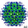
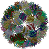
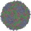
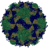
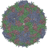
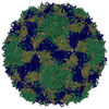
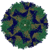
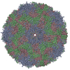
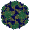
| Title | Engineered Hepatitis B virus core antigen T=3 | |||||||||||||||||||||||||||
 Components Components | Capsid protein,Immunoglobulin G-binding protein A | |||||||||||||||||||||||||||
 Keywords Keywords | VIRUS LIKE PARTICLE / cancer therapy / epidermal growth factor receptor 1 / affibody | |||||||||||||||||||||||||||
| Function / homology |  Function and homology information Function and homology informationmicrotubule-dependent intracellular transport of viral material towards nucleus / IgG binding / T=4 icosahedral viral capsid / viral penetration into host nucleus / host cell / host cell cytoplasm / symbiont entry into host cell / structural molecule activity / DNA binding / RNA binding Similarity search - Function | |||||||||||||||||||||||||||
| Biological species | Hepatitis B virus genotype C subtype adr  Staphylococcus aureus (bacteria) Staphylococcus aureus (bacteria) | |||||||||||||||||||||||||||
| Method | ELECTRON MICROSCOPY / single particle reconstruction / cryo EM / Resolution: 3.6 Å | |||||||||||||||||||||||||||
 Authors Authors | Jeong, H. / Heo, Y. / Yoo, Y. / Ryu, B. / Yun, J. / Cho, H. / Lee, W. | |||||||||||||||||||||||||||
 Citation Citation | Journal: Int J Mol Sci / Year: 2021 Title: Structural and Functional Characterizations of Cancer Targeting Nanoparticles Based on Hepatitis B Virus Capsid. Authors: Yunseok Heo / Hyeongseop Jeong / Youngki Yoo / Ji-Hye Yun / Bumhan Ryu / Young-Je Cha / Bo-Ram Lee / Ye-Eun Jeon / Jongmin Kim / Sojin Jeong / Eunji Jo / Jae-Sung Woo / Jeewon Lee / Hyun-Soo Cho / Weontae Lee /  Abstract: Cancer targeting nanoparticles have been extensively studied, but stable and applicable agents have yet to be developed. Here, we report stable nanoparticles based on hepatitis B core antigen (HBcAg) ...Cancer targeting nanoparticles have been extensively studied, but stable and applicable agents have yet to be developed. Here, we report stable nanoparticles based on hepatitis B core antigen (HBcAg) for cancer therapy. HBcAg monomers assemble into spherical capsids of 180 or 240 subunits. HBcAg was engineered to present an affibody for binding to human epidermal growth factor receptor 1 (EGFR) and to present histidine and tyrosine tags for binding to gold ions. The HBcAg engineered to present affibody and tags (HAF) bound specifically to EGFR and exterminated the EGFR-overexpressing adenocarcinomas under alternating magnetic field (AMF) after binding with gold ions. Using cryogenic electron microscopy (cryo-EM), we obtained the molecular structures of recombinant HAF and found that the overall structure of HAF was the same as that of HBcAg, except with the affibody on the spike. Therefore, HAF is viable for cancer therapy with the advantage of maintaining a stable capsid form. If the affibody in HAF is replaced with a specific sequence to bind to another targetable disease protein, the nanoparticles can be used for drug development over a wide spectrum. | |||||||||||||||||||||||||||
| History |
|
- Structure visualization
| Movie |
Movie viewer |
|---|---|
| Structure viewer | Molecule: MolmilJmol/JSmol |
- Downloads & links
-Download
| PDBx/mmCIF format | 7eoy.cif.gz | 99 KB | Display | PDBx/mmCIF format |
|---|---|---|---|---|
| PDB format | pdb7eoy.ent.gz | 70 KB | Display | PDB format |
| PDBx/mmJSON format | 7eoy.json.gz | Tree view | PDBx/mmJSON format | |
| Others |  Other downloads Other downloads |
-Validation report
| Arichive directory | https://data.pdbj.org/pub/pdb/validation_reports/eo/7eoyftp://data.pdbj.org/pub/pdb/validation_reports/eo/7eoy | HTTPS FTP |
|---|
-Related structure data
| Related structure data |  31233MC  7ep6C  7fdjC M: map data used to model this data C: citing same article ( |
|---|---|
| Similar structure data |


-Links
 PDBj
PDBj


- Assembly
| Deposited unit | 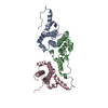
|
|---|---|
| 1 | x 60
|
| 2 |
|
| 3 | x 5
|
| 4 | x 6
|
| 5 | 
|
| Symmetry | Point symmetry: (Schoenflies symbol: I (icosahedral)) |
-Components
| #1: Antibody | Mass: 36803.078 Da / Num. of mol.: 3 Source method: isolated from a genetically manipulated source Details: MHHHHHHMASSLRQILDSQKMEWRSNAGGSGGGSGGGTGGGGGGYYYYYY (expression tag) DIDPYKEFGASVELLSFLPSDFFPSIRDLLDTASALYREALESPEHCSPHHTALRQAILCWGELMNLATWVGSNLED (P69706, residues 2-78) GGGGSGGGGT (linker) ...Details: MHHHHHHMASSLRQILDSQKMEWRSNAGGSGGGSGGGTGGGGGGYYYYYY (expression tag) DIDPYKEFGASVELLSFLPSDFFPSIRDLLDTASALYREALESPEHCSPHHTALRQAILCWGELMNLATWVGSNLED (P69706, residues 2-78) GGGGSGGGGT (linker) LE (enzyme sitelinker) VDNKFNKEMWAAWEEIRNLPNLNGWQMTAFIASLVDDPSQSANLLAEAKKLNDAQAPK (P38507, residues 212-269 => modified) EF (linker) VDNKFNKEMWAAWEEIRNLPNLNGWQMTAFIASLVDDPSQSANLLAEAKKLNDAQAPK (P38507, residues 212-269 => modified) GS (enzyme sitelinker) GGGGSGGGG (linker) SRELVVSYVNVNMGLKIRQLLWFHISCLTFGRETVLEYLVSFGVWIRTPPAYRPPNAPILSTLPETTVV (P69706, residues 81-149) Source: (gene. exp.)  Hepatitis B virus genotype C subtype adr (strain Japan/adr4/1983), (gene. exp.) Staphylococcus aureus (bacteria) Hepatitis B virus genotype C subtype adr (strain Japan/adr4/1983), (gene. exp.) Staphylococcus aureus (bacteria)Gene: spa / Production host: Has protein modification | Y | |
|---|
-Experimental details
-Experiment
| Experiment | Method: ELECTRON MICROSCOPY |
|---|---|
| EM experiment | Aggregation state: PARTICLE / 3D reconstruction method: single particle reconstruction |
- Sample preparation
| Component | Name: Staphylococcus aureus / Type: VIRUS / Details: virus core antigen / Entity ID: all / Source: MULTIPLE SOURCES | ||||||||||||
|---|---|---|---|---|---|---|---|---|---|---|---|---|---|
| Source (natural) |
| ||||||||||||
| Source (recombinant) | Organism: | ||||||||||||
| Details of virus | Empty: YES / Enveloped: NO / Isolate: OTHER / Type: VIRUS-LIKE PARTICLE | ||||||||||||
| Buffer solution | pH: 7.5 | ||||||||||||
| Specimen | Embedding applied: NO / Shadowing applied: NO / Staining applied: NO / Vitrification applied: YES | ||||||||||||
| Specimen support | Grid material: COPPER / Grid mesh size: 200 divisions/in. / Grid type: Quantifoil R1.2/1.3 | ||||||||||||
| Vitrification | Cryogen name: ETHANE |
- Electron microscopy imaging
| Experimental equipment |  Model: Titan Krios / Image courtesy: FEI Company |
|---|---|
| Microscopy | Model: FEI TITAN KRIOS |
| Electron gun | Electron source:  FIELD EMISSION GUN / Accelerating voltage: 300 kV / Illumination mode: FLOOD BEAM FIELD EMISSION GUN / Accelerating voltage: 300 kV / Illumination mode: FLOOD BEAM |
| Electron lens | Mode: BRIGHT FIELD |
| Image recording | Electron dose: 45 e/Å2 / Film or detector model: FEI FALCON III (4k x 4k) |
- Processing
| Software | Name: PHENIX / Version: 1.19.2_4158: / Classification: refinement | ||||||||||||||||||||||||
|---|---|---|---|---|---|---|---|---|---|---|---|---|---|---|---|---|---|---|---|---|---|---|---|---|---|
| EM software | Name: PHENIX / Category: model refinement | ||||||||||||||||||||||||
| CTF correction | Type: PHASE FLIPPING AND AMPLITUDE CORRECTION | ||||||||||||||||||||||||
| 3D reconstruction | Resolution: 3.6 Å / Resolution method: FSC 0.143 CUT-OFF / Num. of particles: 50321 / Symmetry type: POINT | ||||||||||||||||||||||||
| Refine LS restraints |
|
