 Movie
Movie Controller


+ Open data

- Basic information
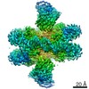
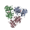
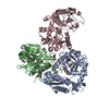
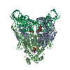
| Entry | Database: PDB / ID: 7dni | |||||||||||||||||||||||||||
|---|---|---|---|---|---|---|---|---|---|---|---|---|---|---|---|---|---|---|---|---|---|---|---|---|---|---|---|---|
| Title | MDA5 CARDs-MAVS CARD polyUb complex | |||||||||||||||||||||||||||
 Components Components |
| |||||||||||||||||||||||||||
 Keywords Keywords | IMMUNE SYSTEM / Signaling / polyubiquitin | |||||||||||||||||||||||||||
| Function / homology |  Function and homology information Function and homology informationpositive regulation of IP-10 production / regulation of peroxisome organization / MDA-5 signaling pathway / regulation of type III interferon production / positive regulation of chemokine (C-C motif) ligand 5 production / positive regulation of myeloid dendritic cell cytokine production / CARD domain binding / detection of virus / positive regulation of response to cytokine stimulus / NF-kB activation through FADD/RIP-1 pathway mediated by caspase-8 and -10 ...positive regulation of IP-10 production / regulation of peroxisome organization / MDA-5 signaling pathway / regulation of type III interferon production / positive regulation of chemokine (C-C motif) ligand 5 production / positive regulation of myeloid dendritic cell cytokine production / CARD domain binding / detection of virus / positive regulation of response to cytokine stimulus / NF-kB activation through FADD/RIP-1 pathway mediated by caspase-8 and -10 / protein localization to mitochondrion / positive regulation of type I interferon-mediated signaling pathway / hypothalamus gonadotrophin-releasing hormone neuron development / Modulation of host responses by IFN-stimulated genes / female meiosis I / positive regulation of protein monoubiquitination / peroxisomal membrane / TRAF6 mediated IRF7 activation / fat pad development / mitochondrion transport along microtubule / negative regulation of viral genome replication / type I interferon-mediated signaling pathway / seminiferous tubule development / cellular response to exogenous dsRNA / pattern recognition receptor activity / female gonad development / cytoplasmic pattern recognition receptor signaling pathway / positive regulation of NLRP3 inflammasome complex assembly / TRAF6 mediated NF-kB activation / protein complex oligomerization / male meiosis I / positive regulation of interferon-alpha production / protein sumoylation / positive regulation of intrinsic apoptotic signaling pathway by p53 class mediator / ribonucleoprotein complex binding / ubiquitin ligase complex / neuron projection morphogenesis / cellular response to interferon-beta / positive regulation of defense response to virus by host / energy homeostasis / positive regulation of type I interferon production / regulation of proteasomal protein catabolic process / Maturation of protein E / Maturation of protein E / ER Quality Control Compartment (ERQC) / signaling adaptor activity / Myoclonic epilepsy of Lafora / FLT3 signaling by CBL mutants / activation of innate immune response / IRAK2 mediated activation of TAK1 complex / Prevention of phagosomal-lysosomal fusion / Alpha-protein kinase 1 signaling pathway / Glycogen synthesis / IRAK1 recruits IKK complex / IRAK1 recruits IKK complex upon TLR7/8 or 9 stimulation / Endosomal Sorting Complex Required For Transport (ESCRT) / Membrane binding and targetting of GAG proteins / Negative regulation of FLT3 / Regulation of TBK1, IKKε (IKBKE)-mediated activation of IRF3, IRF7 / PTK6 Regulates RTKs and Their Effectors AKT1 and DOK1 / positive regulation of interferon-beta production / Regulation of TBK1, IKKε-mediated activation of IRF3, IRF7 upon TLR3 ligation / IRAK2 mediated activation of TAK1 complex upon TLR7/8 or 9 stimulation / Constitutive Signaling by NOTCH1 HD Domain Mutants / NOTCH2 Activation and Transmission of Signal to the Nucleus / TICAM1,TRAF6-dependent induction of TAK1 complex / TICAM1-dependent activation of IRF3/IRF7 / APC/C:Cdc20 mediated degradation of Cyclin B / Regulation of FZD by ubiquitination / Downregulation of ERBB4 signaling / APC-Cdc20 mediated degradation of Nek2A / p75NTR recruits signalling complexes / InlA-mediated entry of Listeria monocytogenes into host cells / TRAF6 mediated IRF7 activation in TLR7/8 or 9 signaling / regulation of neuron apoptotic process / NF-kB is activated and signals survival / antiviral innate immune response / TRAF6-mediated induction of TAK1 complex within TLR4 complex / Regulation of pyruvate metabolism / Pexophagy / Regulation of innate immune responses to cytosolic DNA / NRIF signals cell death from the nucleus / Downregulation of ERBB2:ERBB3 signaling / Regulation of PTEN localization / VLDLR internalisation and degradation / positive regulation of protein ubiquitination / Activated NOTCH1 Transmits Signal to the Nucleus / Synthesis of active ubiquitin: roles of E1 and E2 enzymes / Translesion synthesis by REV1 / TICAM1, RIP1-mediated IKK complex recruitment / Regulation of BACH1 activity / Translesion synthesis by POLK / InlB-mediated entry of Listeria monocytogenes into host cell / JNK (c-Jun kinases) phosphorylation and activation mediated by activated human TAK1 / Activation of IRF3, IRF7 mediated by TBK1, IKKε (IKBKE) / MAP3K8 (TPL2)-dependent MAPK1/3 activation / Downregulation of TGF-beta receptor signaling / Translesion synthesis by POLI / regulation of mitochondrial membrane potential / positive regulation of interleukin-8 production Similarity search - Function | |||||||||||||||||||||||||||
| Biological species |  Homo sapiens (human) Homo sapiens (human) | |||||||||||||||||||||||||||
| Method | ELECTRON MICROSCOPY / single particle reconstruction / Resolution: 3.2 Å | |||||||||||||||||||||||||||
 Authors Authors | Song, B. / Chen, Y. / Luo, D.H. / Zheng, J. | |||||||||||||||||||||||||||
| Funding support |  China, 1items China, 1items
| |||||||||||||||||||||||||||
 Citation Citation | Journal: Immunity / Year: 2021 Title: Ordered assembly of the cytosolic RNA-sensing MDA5-MAVS signaling complex via binding to unanchored K63-linked poly-ubiquitin chains. Authors: Bin Song / Yun Chen / Xin Liu / Fei Yuan / Eddie Yong Jun Tan / Yixuan Lei / Ning Song / Yinqi Han / Bruce D Pascal / Patrick R Griffin / Cheng Luo / Bin Wu / Dahai Luo / Jie Zheng /   Abstract: The RNA sensor MDA5 recruits the signaling adaptor MAVS to initiate type I interferon signaling and downstream antiviral responses, a process that requires K63-linked polyubiquitin chains. Here, we ...The RNA sensor MDA5 recruits the signaling adaptor MAVS to initiate type I interferon signaling and downstream antiviral responses, a process that requires K63-linked polyubiquitin chains. Here, we examined the mechanisms whereby K63-polyUb chain regulate MDA5 activation. Only long unanchored K63-polyUb (n ≥ 8) could mediate tetramerization of the caspase activation and recruitment domains of MDA5 (CARDs). Cryoelectron microscopy structures of a polyUb-bound CARDs tetramer and a polyUb-bound CARDs-CARD assembly revealed a tower-like formation, wherein eight Ubs tethered along the outer rim of the helical shell, bridging CARDs and CARD tetramers into proximity. ATP binding and hydrolysis promoted the stabilization of RNA-bound MDA5 prior to MAVS activation via allosteric effects on CARDs-polyUb complex. Abundant ATP prevented basal activation of apo MDA5. Our findings reveal the ordered assembly of a MDA5 signaling complex competent to recruit and activate MAVS and highlight differences with RIG-I in terms of CARD orientation and Ub sensing that suggest different abilities to induce antiviral responses. | |||||||||||||||||||||||||||
| History |
|
- Structure visualization
| Movie |
Movie viewer |
|---|---|
| Structure viewer | Molecule: MolmilJmol/JSmol |
- Downloads & links
-Download
| PDBx/mmCIF format | 7dni.cif.gz | 307.8 KB | Display | PDBx/mmCIF format |
|---|---|---|---|---|
| PDB format | pdb7dni.ent.gz | 252.7 KB | Display | PDB format |
| PDBx/mmJSON format | 7dni.json.gz | Tree view | PDBx/mmJSON format | |
| Others |  Other downloads Other downloads |
-Validation report
| Arichive directory | https://data.pdbj.org/pub/pdb/validation_reports/dn/7dniftp://data.pdbj.org/pub/pdb/validation_reports/dn/7dni | HTTPS FTP |
|---|
-Related structure data
| Related structure data |  30784MC  7dnjC M: map data used to model this data C: citing same article ( |
|---|---|
| Similar structure data |

-Links
 PDBj
PDBj


- Assembly
| Deposited unit | 
|
|---|---|
| 1 |
|
-Components
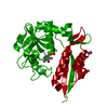
| #1: Protein | Mass: 8576.831 Da / Num. of mol.: 8 Source method: isolated from a genetically manipulated source Source: (gene. exp.) Homo sapiens (human) / Gene: UBB / Production host:  #2: Protein | Mass: 23825.883 Da / Num. of mol.: 4 Source method: isolated from a genetically manipulated source Source: (gene. exp.) Homo sapiens (human) / Gene: MDA5 / Production host: #3: Protein | Mass: 11342.082 Da / Num. of mol.: 4 / Mutation: D23K, E26K, E80K Source method: isolated from a genetically manipulated source Source: (gene. exp.) Homo sapiens (human) / Gene: MAVS / Production host: Has protein modification | N | |
|---|
-Experimental details
-Experiment
| Experiment | Method: ELECTRON MICROSCOPY |
|---|---|
| EM experiment | Aggregation state: PARTICLE / 3D reconstruction method: single particle reconstruction |
- Sample preparation
| Component | Name: K63-polyUb bound MDA5CARDs-MAVSCARD complex / Type: COMPLEX / Entity ID: all / Source: RECOMBINANT |
|---|---|
| Source (natural) | Organism: Homo sapiens (human) |
| Source (recombinant) | Organism: |
| Buffer solution | pH: 7.5 |
| Specimen | Conc.: 1.6 mg/ml / Embedding applied: NO / Shadowing applied: NO / Staining applied: NO / Vitrification applied: NO / Details: heterotetrameric complex |
- Electron microscopy imaging
| Experimental equipment |  Model: Titan Krios / Image courtesy: FEI Company |
|---|---|
| Microscopy | Model: FEI TITAN KRIOS |
| Electron gun | Electron source:  FIELD EMISSION GUN / Accelerating voltage: 300 kV / Illumination mode: OTHER FIELD EMISSION GUN / Accelerating voltage: 300 kV / Illumination mode: OTHER |
| Electron lens | Mode: BRIGHT FIELD |
| Image recording | Electron dose: 70 e/Å2 / Film or detector model: GATAN K3 BIOQUANTUM (6k x 4k) |
- Processing
| Software | Name: PHENIX / Version: 1.18.2_3874: / Classification: refinement | ||||||||||||||||||||||||
|---|---|---|---|---|---|---|---|---|---|---|---|---|---|---|---|---|---|---|---|---|---|---|---|---|---|
| EM software | Name: PHENIX / Category: model refinement | ||||||||||||||||||||||||
| CTF correction | Type: NONE | ||||||||||||||||||||||||
| 3D reconstruction | Resolution: 3.2 Å / Resolution method: FSC 0.143 CUT-OFF / Num. of particles: 212112 / Symmetry type: POINT | ||||||||||||||||||||||||
| Refine LS restraints |
|
