 Movie
Movie Controller

[English] 日本語
 Yorodumi
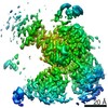
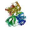
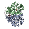
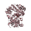
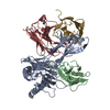
- PDB-7dcr: cryo-EM structure of the DEAH-box helicase Prp2 in complex with i... -
+ Open data

- Basic information
| Entry | Database: PDB / ID: 7dcr | |||||||||||||||||||||||||||
|---|---|---|---|---|---|---|---|---|---|---|---|---|---|---|---|---|---|---|---|---|---|---|---|---|---|---|---|---|
| Title | cryo-EM structure of the DEAH-box helicase Prp2 in complex with its coactivator Spp2 | |||||||||||||||||||||||||||
 Components Components |
| |||||||||||||||||||||||||||
 Keywords Keywords | SPLICING / spliceosome / RNA splicing / Bact complex / DEAH-box ATPase/helicase / Prp2 / Spp2 | |||||||||||||||||||||||||||
| Function / homology |  Function and homology information Function and homology informationsnoRNA splicing / generation of catalytic spliceosome for first transesterification step / ATP-dependent activity, acting on RNA / U2-type catalytic step 1 spliceosome / ATPase activator activity / Prp19 complex / spliceosomal complex / helicase activity / mRNA splicing, via spliceosome / nucleic acid binding ...snoRNA splicing / generation of catalytic spliceosome for first transesterification step / ATP-dependent activity, acting on RNA / U2-type catalytic step 1 spliceosome / ATPase activator activity / Prp19 complex / spliceosomal complex / helicase activity / mRNA splicing, via spliceosome / nucleic acid binding / RNA helicase activity / RNA helicase / ATP hydrolysis activity / RNA binding / ATP binding / nucleus / cytoplasm Similarity search - Function | |||||||||||||||||||||||||||
| Biological species |  | |||||||||||||||||||||||||||
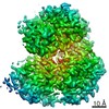
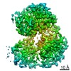
| Method | ELECTRON MICROSCOPY / single particle reconstruction / cryo EM / Resolution: 3.15 Å | |||||||||||||||||||||||||||
 Authors Authors | Bai, R. / Wan, R. / Yan, C. / Jia, Q. / Zhang, P. / Lei, J. / Shi, Y. | |||||||||||||||||||||||||||
| Funding support |  China, 1items China, 1items
| |||||||||||||||||||||||||||
 Citation Citation | Journal: Science / Year: 2021 Title: Mechanism of spliceosome remodeling by the ATPase/helicase Prp2 and its coactivator Spp2. Authors: Rui Bai / Ruixue Wan / Chuangye Yan / Qi Jia / Jianlin Lei / Yigong Shi / Abstract: Spliceosome remodeling, executed by conserved adenosine triphosphatase (ATPase)/helicases including Prp2, enables precursor messenger RNA (pre-mRNA) splicing. However, the structural basis for the ...Spliceosome remodeling, executed by conserved adenosine triphosphatase (ATPase)/helicases including Prp2, enables precursor messenger RNA (pre-mRNA) splicing. However, the structural basis for the function of the ATPase/helicases remains poorly understood. Here, we report atomic structures of Prp2 in isolation, Prp2 complexed with its coactivator Spp2, and Prp2-loaded activated spliceosome and the results of structure-guided biochemical analysis. Prp2 weakly associates with the spliceosome and cannot function without Spp2, which stably associates with Prp2 and anchors on the spliceosome, thus tethering Prp2 to the activated spliceosome and allowing Prp2 to function. Pre-mRNA is loaded into a featured channel between the N and C halves of Prp2, where Leu from the N half and Arg from the C half prevent backward sliding of pre-mRNA toward its 5'-end. Adenosine 5'-triphosphate binding and hydrolysis trigger interdomain movement in Prp2, which drives unidirectional stepwise translocation of pre-mRNA toward its 3'-end. These conserved mechanisms explain the coupling of spliceosome remodeling to pre-mRNA splicing. | |||||||||||||||||||||||||||
| History |
|
- Structure visualization
| Movie |
Movie viewer |
|---|---|
| Structure viewer | Molecule: MolmilJmol/JSmol |
- Downloads & links
-Download
| PDBx/mmCIF format | 7dcr.cif.gz | 142.9 KB | Display | PDBx/mmCIF format |
|---|---|---|---|---|
| PDB format | pdb7dcr.ent.gz | 105.9 KB | Display | PDB format |
| PDBx/mmJSON format | 7dcr.json.gz | Tree view | PDBx/mmJSON format | |
| Others |  Other downloads Other downloads |
-Validation report
| Summary document | 7dcr_validation.pdf.gz | 889.4 KB | Display | wwPDB validaton report |
|---|---|---|---|---|
| Full document | 7dcr_full_validation.pdf.gz | 892.5 KB | Display | |
| Data in XML | 7dcr_validation.xml.gz | 24.9 KB | Display | |
| Data in CIF | 7dcr_validation.cif.gz | 36.9 KB | Display | |
| Arichive directory | https://data.pdbj.org/pub/pdb/validation_reports/dc/7dcrftp://data.pdbj.org/pub/pdb/validation_reports/dc/7dcr | HTTPS FTP |
-Related structure data
| Related structure data | 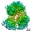 30640MC 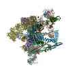 7dcoC 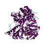 7dcpC 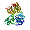 7dcqC 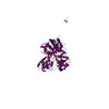 7dd3C M: map data used to model this data C: citing same article ( |
|---|---|
| Similar structure data |


-Links
 PDBj
PDBj

- Assembly
| Deposited unit | 
|
|---|---|
| 1 |
|
-Components
| #1: Protein | Mass: 99947.492 Da / Num. of mol.: 1 Source method: isolated from a genetically manipulated source Source: (gene. exp.) Gene: PRP2, GI526_G0005089, PACBIOSEQ_LOCUS5743 / Production host:  |
|---|---|
| #2: Protein | Mass: 20685.377 Da / Num. of mol.: 1 Source method: isolated from a genetically manipulated source Source: (gene. exp.) Gene: SPP2, GI526_G0005483 / Production host: |
| Has protein modification | N |
-Experimental details
-Experiment
| Experiment | Method: ELECTRON MICROSCOPY |
|---|---|
| EM experiment | Aggregation state: PARTICLE / 3D reconstruction method: single particle reconstruction |
- Sample preparation
| Component | Name: Complex of ATPase/helicase Prp2 and its coactivator Spp2 Type: COMPLEX / Entity ID: all / Source: RECOMBINANT |
|---|---|
| Molecular weight | Value: 119 kDa/nm / Experimental value: YES |
| Source (natural) | Organism: |
| Source (recombinant) | Organism: |
| Buffer solution | pH: 7.9 |
| Specimen | Embedding applied: NO / Shadowing applied: NO / Staining applied: NO / Vitrification applied: YES |
| Specimen support | Grid material: GOLD / Grid type: Quantifoil R1.2/1.3 |
| Vitrification | Cryogen name: ETHANE / Humidity: 100 % |
- Electron microscopy imaging
| Microscopy | Model: FEI TITAN |
|---|---|
| Electron gun | Electron source:  FIELD EMISSION GUN / Accelerating voltage: 300 kV / Illumination mode: FLOOD BEAM FIELD EMISSION GUN / Accelerating voltage: 300 kV / Illumination mode: FLOOD BEAM |
| Electron lens | Mode: BRIGHT FIELD |
| Image recording | Electron dose: 50 e/Å2 / Film or detector model: GATAN K3 (6k x 4k) |
| EM imaging optics | Energyfilter slit width: 20 eV |
- Processing
| Software | Name: PHENIX / Version: 1.13_2998: / Classification: refinement |
|---|---|
| EM software | Name: PHENIX / Category: model refinement |
| CTF correction | Type: PHASE FLIPPING AND AMPLITUDE CORRECTION |
| 3D reconstruction | Resolution: 3.15 Å / Resolution method: FSC 0.143 CUT-OFF / Num. of particles: 130302 / Symmetry type: POINT |
