 Movie
Movie Controller


+ Open data

- Basic information
| Entry | Database: PDB / ID: 7b5f | ||||||
|---|---|---|---|---|---|---|---|
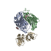
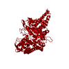
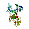
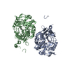
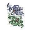
| Title | Structure of echovirus 18 in complex with neonatal Fc receptor | ||||||
 Components Components |
| ||||||
 Keywords Keywords | VIRUS / complex / echovirus 18 virion / neonatal fc receptor / fcrn / beta-2-microglobulin / microglobulin / pocket factor | ||||||
| Function / homology |  Function and homology information Function and homology informationIgG immunoglobulin transcytosis in epithelial cells mediated by FcRn immunoglobulin receptor / IgG binding / symbiont-mediated suppression of host cytoplasmic pattern recognition receptor signaling pathway via inhibition of RIG-I activity / beta-2-microglobulin binding / early endosome lumen / Nef mediated downregulation of MHC class I complex cell surface expression / DAP12 interactions / Endosomal/Vacuolar pathway / T cell mediated cytotoxicity / picornain 2A ...IgG immunoglobulin transcytosis in epithelial cells mediated by FcRn immunoglobulin receptor / IgG binding / symbiont-mediated suppression of host cytoplasmic pattern recognition receptor signaling pathway via inhibition of RIG-I activity / beta-2-microglobulin binding / early endosome lumen / Nef mediated downregulation of MHC class I complex cell surface expression / DAP12 interactions / Endosomal/Vacuolar pathway / T cell mediated cytotoxicity / picornain 2A / Antigen Presentation: Folding, assembly and peptide loading of class I MHC / symbiont-mediated suppression of host mRNA export from nucleus / regulation of iron ion transport / cellular response to iron(III) ion / negative regulation of iron ion transport / negative regulation of forebrain neuron differentiation / antigen processing and presentation of exogenous protein antigen via MHC class Ib, TAP-dependent / symbiont genome entry into host cell via pore formation in plasma membrane / picornain 3C / peptide antigen assembly with MHC class I protein complex / ER to Golgi transport vesicle membrane / regulation of erythrocyte differentiation / response to molecule of bacterial origin / HFE-transferrin receptor complex / MHC class I peptide loading complex / transferrin transport / cellular response to iron ion / negative regulation of receptor-mediated endocytosis / T=pseudo3 icosahedral viral capsid / positive regulation of T cell cytokine production / antigen processing and presentation of endogenous peptide antigen via MHC class I / MHC class I protein complex / peptide antigen assembly with MHC class II protein complex / negative regulation of neurogenesis / MHC class II protein complex / cellular response to nicotine / positive regulation of receptor-mediated endocytosis / multicellular organismal-level iron ion homeostasis / host cell cytoplasmic vesicle membrane / positive regulation of T cell mediated cytotoxicity / specific granule lumen / antigen processing and presentation of exogenous peptide antigen via MHC class II / positive regulation of immune response / peptide antigen binding / phagocytic vesicle membrane / recycling endosome membrane / positive regulation of T cell activation / negative regulation of epithelial cell proliferation / Interferon gamma signaling / Immunoregulatory interactions between a Lymphoid and a non-Lymphoid cell / sensory perception of smell / Modulation by Mtb of host immune system / positive regulation of cellular senescence / tertiary granule lumen / MHC class II protein complex binding / ribonucleoside triphosphate phosphatase activity / T cell differentiation in thymus / DAP12 signaling / late endosome membrane / negative regulation of neuron projection development / nucleoside-triphosphate phosphatase / protein refolding / channel activity / ER-Phagosome pathway / early endosome membrane / monoatomic ion transmembrane transport / amyloid fibril formation / protein homotetramerization / intracellular iron ion homeostasis / learning or memory / DNA replication / RNA helicase activity / endosome membrane / immune response / endocytosis involved in viral entry into host cell / endoplasmic reticulum lumen / Amyloid fiber formation / Golgi membrane / external side of plasma membrane / symbiont-mediated activation of host autophagy / RNA-directed RNA polymerase / lysosomal membrane / cysteine-type endopeptidase activity / focal adhesion / viral RNA genome replication / RNA-directed RNA polymerase activity / Neutrophil degranulation / virion attachment to host cell / host cell nucleus / DNA-templated transcription / SARS-CoV-2 activates/modulates innate and adaptive immune responses / structural molecule activity / Golgi apparatus / endoplasmic reticulum / protein homodimerization activity / proteolysis / : / RNA binding / extracellular exosome / extracellular region Similarity search - Function | ||||||
| Biological species |  Homo sapiens (human) Homo sapiens (human) Echovirus E18 Echovirus E18 | ||||||
| Method | ELECTRON MICROSCOPY / single particle reconstruction / cryo EM / Resolution: 2.9 Å | ||||||
 Authors Authors | Buchta, D. / Levdansky, Y. / Fuzik, T. / Mukhamedova, L. / Moravcova, J. / Hrebik, D. / Andersen, J.T. / Plevka, P. | ||||||
| Funding support |  Czech Republic, 1items Czech Republic, 1items
| ||||||
 Citation Citation | Journal: To Be Published Title: Structure of echovirus 18 in complex with neonatal Fc receptor Authors: Buchta, D. / Levdansky, Y. / Fuzik, T. / Mukhamedova, L. / Moravcova, J. / Hrebik, D. / Andersen, J.T. / Plevka, P. | ||||||
| History |
|
- Structure visualization
| Movie |
Movie viewer |
|---|---|
| Structure viewer | Molecule: MolmilJmol/JSmol |
- Downloads & links
-Download
| PDBx/mmCIF format | 7b5f.cif.gz | 185.6 KB | Display | PDBx/mmCIF format |
|---|---|---|---|---|
| PDB format | pdb7b5f.ent.gz | 140.7 KB | Display | PDB format |
| PDBx/mmJSON format | 7b5f.json.gz | Tree view | PDBx/mmJSON format | |
| Others |  Other downloads Other downloads |
-Validation report
| Arichive directory | https://data.pdbj.org/pub/pdb/validation_reports/b5/7b5fftp://data.pdbj.org/pub/pdb/validation_reports/b5/7b5f | HTTPS FTP |
|---|
-Related structure data
| Related structure data |  12028MC M: map data used to model this data C: citing same article ( |
|---|---|
| Similar structure data |


-Links
 PDBj
PDBj


- Assembly
| Deposited unit | 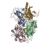
|
|---|---|
| 1 |
|
-Components
-Echovirus 18 viral protein ... , 4 types, 4 molecules CDAB
| #1: Protein | Mass: 26143.783 Da / Num. of mol.: 1 / Source method: isolated from a natural source / Source: (natural) Echovirus E18 / Cell line: GMK / Tissue: KidneyReferences: UniProt: Q8V635, picornain 2A, nucleoside-triphosphate phosphatase, picornain 3C, RNA-directed RNA polymerase |
|---|---|
| #2: Protein | Mass: 7475.322 Da / Num. of mol.: 1 / Source method: isolated from a natural source / Source: (natural) Echovirus E18 / Cell line: GMK / Organ: KidneyReferences: UniProt: Q8V635, picornain 2A, nucleoside-triphosphate phosphatase, picornain 3C, RNA-directed RNA polymerase |
| #3: Protein | Mass: 32564.445 Da / Num. of mol.: 1 / Source method: isolated from a natural source / Source: (natural) Echovirus E18 / Cell line: GMK / Tissue: KidneyReferences: UniProt: Q8V635, picornain 2A, nucleoside-triphosphate phosphatase, picornain 3C, RNA-directed RNA polymerase |
| #4: Protein | Mass: 28802.328 Da / Num. of mol.: 1 / Source method: isolated from a natural source / Source: (natural) Echovirus E18 / Cell line: GMK / Tissue: KidneyReferences: UniProt: Q8V635, picornain 2A, nucleoside-triphosphate phosphatase, picornain 3C, RNA-directed RNA polymerase |
-Protein , 2 types, 2 molecules GH
| #5: Protein | Mass: 29720.383 Da / Num. of mol.: 1 Source method: isolated from a genetically manipulated source Source: (gene. exp.) Homo sapiens (human) / Gene: FCGRT, FCRN / Cell line (production host): High Five cells / Production host: unidentified baculovirus / References: UniProt: P55899 |
|---|---|
| #6: Protein | Mass: 11748.160 Da / Num. of mol.: 1 Source method: isolated from a genetically manipulated source Source: (gene. exp.) Homo sapiens (human) / Gene: B2M, CDABP0092, HDCMA22P / Cell line (production host): High Five cells / Production host: unidentified baculovirus / References: UniProt: P61769 |
-Non-polymers , 2 types, 2 molecules 
| #7: Chemical | ChemComp-PLM /  Mass: 256.424 Da / Num. of mol.: 1 / Source method: obtained synthetically / Formula: C16H32O2 / Feature type: SUBJECT OF INVESTIGATION Mass: 256.424 Da / Num. of mol.: 1 / Source method: obtained synthetically / Formula: C16H32O2 / Feature type: SUBJECT OF INVESTIGATION |
|---|---|
| #8: Chemical | ChemComp-GUN /  Mass: 151.126 Da / Num. of mol.: 1 / Source method: obtained synthetically / Formula: C5H5N5O Mass: 151.126 Da / Num. of mol.: 1 / Source method: obtained synthetically / Formula: C5H5N5O |
-Details
| Has ligand of interest | Y |
|---|
-Experimental details
-Experiment
| Experiment | Method: ELECTRON MICROSCOPY |
|---|---|
| EM experiment | Aggregation state: PARTICLE / 3D reconstruction method: single particle reconstruction |
- Sample preparation
| Component |
| ||||||||||||||||||||||||||||||
|---|---|---|---|---|---|---|---|---|---|---|---|---|---|---|---|---|---|---|---|---|---|---|---|---|---|---|---|---|---|---|---|
| Molecular weight |
| ||||||||||||||||||||||||||||||
| Source (natural) |
| ||||||||||||||||||||||||||||||
| Source (recombinant) | Organism: unidentified baculovirus | ||||||||||||||||||||||||||||||
| Natural host | Organism: homo sapiens / Strain: echovirus 18 | ||||||||||||||||||||||||||||||
| Buffer solution | pH: 7.3 Details: Mixed in 1:1 volume ratio {20 mM Tris (pH=7.2), 100 mM NaCl} and {8 mM Na2HPO4, 2 mM KH2PO4 (pH=7.4), 137 mM NaCl, 2.7 mM KCl}=PBS | ||||||||||||||||||||||||||||||
| Buffer component |
| ||||||||||||||||||||||||||||||
| Specimen | Conc.: 2 mg/ml / Embedding applied: NO / Shadowing applied: NO / Staining applied: NO / Vitrification applied: YES | ||||||||||||||||||||||||||||||
| Specimen support | Grid material: COPPER / Grid mesh size: 300 divisions/in. / Grid type: Quantifoil R2/1 | ||||||||||||||||||||||||||||||
| Vitrification | Instrument: FEI VITROBOT MARK IV / Cryogen name: ETHANE |
- Electron microscopy imaging
| Experimental equipment |  Model: Titan Krios / Image courtesy: FEI Company |
|---|---|
| Microscopy | Model: FEI TITAN KRIOS |
| Electron gun | Electron source:  FIELD EMISSION GUN / Accelerating voltage: 300 kV / Illumination mode: FLOOD BEAM FIELD EMISSION GUN / Accelerating voltage: 300 kV / Illumination mode: FLOOD BEAM |
| Electron lens | Mode: BRIGHT FIELD / Nominal magnification: 75000 X / Nominal defocus max: 2400 nm / Nominal defocus min: 300 nm / Cs: 2.7 mm / C2 aperture diameter: 50 µm / Alignment procedure: COMA FREE |
| Specimen holder | Cryogen: NITROGEN / Specimen holder model: FEI TITAN KRIOS AUTOGRID HOLDER |
| Image recording | Average exposure time: 1.27 sec. / Electron dose: 54.48 e/Å2 / Detector mode: INTEGRATING / Film or detector model: FEI FALCON III (4k x 4k) / Num. of real images: 4531 / Details: Images were collected in movie-mode at 50 frames. |
| Image scans | Sampling size: 14 µm / Width: 4096 / Height: 4096 |
- Processing
| Software | Name: PHENIX / Version: (1.13_2992: ???) / Classification: refinement | ||||||||||||||||||||||||
|---|---|---|---|---|---|---|---|---|---|---|---|---|---|---|---|---|---|---|---|---|---|---|---|---|---|
| EM software |
| ||||||||||||||||||||||||
| CTF correction | Type: PHASE FLIPPING ONLY | ||||||||||||||||||||||||
| Particle selection | Num. of particles selected: 125031 | ||||||||||||||||||||||||
| Symmetry | Point symmetry: I (icosahedral) | ||||||||||||||||||||||||
| 3D reconstruction | Resolution: 2.9 Å / Resolution method: FSC 0.143 CUT-OFF / Num. of particles: 13062 / Algorithm: FOURIER SPACE / Symmetry type: POINT | ||||||||||||||||||||||||
| Atomic model building | Method: flexible fit / Target criteria: R-factors | ||||||||||||||||||||||||
| Refinement | Resolution: 2.9→233.42 Å / SU ML: 0.65 / σ(F): 0.13 / Phase error: 34.06 / Stereochemistry target values: ML
| ||||||||||||||||||||||||
| Solvent computation | Shrinkage radii: 0.9 Å / VDW probe radii: 1.11 Å / Solvent model: FLAT BULK SOLVENT MODEL | ||||||||||||||||||||||||
| Refine LS restraints |
|
