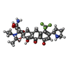
+ Open data
- Basic information
| Entry | Database: PDB / ID: 7ry3 | ||||||
|---|---|---|---|---|---|---|---|
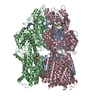
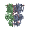
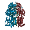
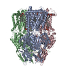
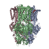
| Title | Multidrug Efflux pump AdeJ with TP-6076 bound | ||||||
 Components Components | Efflux pump membrane transporter | ||||||
 Keywords Keywords | MEMBRANE PROTEIN/ANTIBIOTIC / multidrug efflux pump / MEMBRANE PROTEIN-ANTIBIOTIC complex | ||||||
| Function / homology |  Function and homology information Function and homology informationefflux transmembrane transporter activity / xenobiotic transmembrane transporter activity / response to toxic substance / plasma membrane Similarity search - Function | ||||||
| Biological species |  Acinetobacter baumannii (bacteria) Acinetobacter baumannii (bacteria) | ||||||
| Method | ELECTRON MICROSCOPY / single particle reconstruction / cryo EM / Resolution: 2.91 Å | ||||||
 Authors Authors | Zhang, Z. | ||||||
| Funding support |  United States, 1items United States, 1items
| ||||||
 Citation Citation |  Journal: mBio / Year: 2021 Journal: mBio / Year: 2021Title: An Analysis of the Novel Fluorocycline TP-6076 Bound to Both the Ribosome and Multidrug Efflux Pump AdeJ from Acinetobacter baumannii. Authors: Christopher E Morgan / Zhemin Zhang / Robert A Bonomo / Edward W Yu /  Abstract: Antibiotic resistance among bacterial pathogens continues to pose a serious global health threat. Multidrug-resistant (MDR) strains of the Gram-negative organism Acinetobacter baumannii utilize a ...Antibiotic resistance among bacterial pathogens continues to pose a serious global health threat. Multidrug-resistant (MDR) strains of the Gram-negative organism Acinetobacter baumannii utilize a number of resistance determinants to evade current antibiotics. One of the major resistance mechanisms employed by these pathogens is the use of multidrug efflux pumps. These pumps extrude xenobiotics directly out of bacterial cells, resulting in treatment failures when common antibiotics are administered. Here, the structure of the novel tetracycline antibiotic TP-6076, bound to both the cinetobacter rug fflux pump AdeJ and the ribosome from Acinetobacter baumannii, using single-particle cryo-electron microscopy (cryo-EM), is elucidated. In this work, the structure of the AdeJ-TP-6076 complex is solved, and we show that AdeJ utilizes a network of hydrophobic interactions to recognize this fluorocycline. Concomitant with this, we elucidate three structures of TP-6076 bound to the A. baumannii ribosome and determine that its binding is stabilized largely by electrostatic interactions. We then compare the differences in binding modes between TP-6076 and the related tetracycline antibiotic eravacycline in both targets. These differences suggest that modifications to the tetracycline core may be able to alter AdeJ binding while maintaining interactions with the ribosome. Together, this work highlights how different mechanisms are used to stabilize the binding of tetracycline-based compounds to unique bacterial targets and provides guidance for the future clinical development of tetracycline antibiotics. Treatment of antibiotic-resistant organisms such as A. baumannii represents an ongoing issue for modern medicine. The multidrug efflux pump AdeJ serves as a major resistance determinant in A. baumannii through its action of extruding antibiotics from the cell. In this work, we use cryo-EM to show how AdeJ recognizes the experimental tetracycline antibiotic TP-6076 and prevents this drug from interacting with the A. baumannii ribosome. Since AdeJ and the ribosome use different binding modes to stabilize interactions with TP-6076, exploiting these differences may guide future drug development for combating antibiotic-resistant A. baumannii and potentially other strains of MDR bacteria. | ||||||
| History |
|
- Structure visualization
| Movie |
 Movie viewer Movie viewer |
|---|---|
| Structure viewer | Molecule:  Molmil Molmil Jmol/JSmol Jmol/JSmol |
- Downloads & links
- Download
| PDBx/mmCIF format |  7ry3.cif.gz 7ry3.cif.gz | 530.9 KB | Display |  PDBx/mmCIF format PDBx/mmCIF format |
|---|---|---|---|---|
| PDB format |  pdb7ry3.ent.gz pdb7ry3.ent.gz | 428.9 KB | Display |  PDB format PDB format |
| PDBx/mmJSON format |  7ry3.json.gz 7ry3.json.gz | Tree view |  PDBx/mmJSON format PDBx/mmJSON format | |
| Others |  Other downloads Other downloads |
-Validation report
| Arichive directory |  https://data.pdbj.org/pub/pdb/validation_reports/ry/7ry3 https://data.pdbj.org/pub/pdb/validation_reports/ry/7ry3 ftp://data.pdbj.org/pub/pdb/validation_reports/ry/7ry3 ftp://data.pdbj.org/pub/pdb/validation_reports/ry/7ry3 | HTTPS FTP |
|---|
-Related structure data
| Related structure data |  24732MC  7ryfC  7rygC  7ryhC M: map data used to model this data C: citing same article ( |
|---|---|
| Similar structure data |
- Links
Links
- Assembly
| Deposited unit | 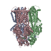
|
|---|---|
| 1 |
|
- Components
| #1: Protein | Mass: 114637.781 Da / Num. of mol.: 3 Source method: isolated from a genetically manipulated source Source: (gene. exp.)  Acinetobacter baumannii (bacteria) / Gene: adeJ, mexB_1, mexB_2 / Production host: Acinetobacter baumannii (bacteria) / Gene: adeJ, mexB_1, mexB_2 / Production host:  #2: Chemical | ChemComp-80P / ( | Has ligand of interest | Y | |
|---|
-Experimental details
-Experiment
| Experiment | Method: ELECTRON MICROSCOPY |
|---|---|
| EM experiment | Aggregation state: PARTICLE / 3D reconstruction method: single particle reconstruction |
- Sample preparation
| Component | Name: multidrug efflux pump AdeJ / Type: COMPLEX / Entity ID: #1 / Source: RECOMBINANT |
|---|---|
| Molecular weight | Experimental value: NO |
| Source (natural) | Organism:  Acinetobacter baumannii (bacteria) Acinetobacter baumannii (bacteria) |
| Source (recombinant) | Organism:  |
| Buffer solution | pH: 7.5 |
| Specimen | Embedding applied: NO / Shadowing applied: NO / Staining applied: NO / Vitrification applied: YES |
| Vitrification | Cryogen name: ETHANE |
- Electron microscopy imaging
| Experimental equipment |  Model: Titan Krios / Image courtesy: FEI Company |
|---|---|
| Microscopy | Model: FEI TITAN KRIOS |
| Electron gun | Electron source:  FIELD EMISSION GUN / Accelerating voltage: 300 kV / Illumination mode: FLOOD BEAM FIELD EMISSION GUN / Accelerating voltage: 300 kV / Illumination mode: FLOOD BEAM |
| Electron lens | Mode: BRIGHT FIELD |
| Image recording | Electron dose: 36 e/Å2 / Film or detector model: GATAN K3 BIOQUANTUM (6k x 4k) |
- Processing
| Software |
| ||||||||||||||||||||||||
|---|---|---|---|---|---|---|---|---|---|---|---|---|---|---|---|---|---|---|---|---|---|---|---|---|---|
| CTF correction | Type: PHASE FLIPPING AND AMPLITUDE CORRECTION | ||||||||||||||||||||||||
| 3D reconstruction | Resolution: 2.91 Å / Resolution method: FSC 0.143 CUT-OFF / Num. of particles: 68346 / Symmetry type: POINT | ||||||||||||||||||||||||
| Refinement | Cross valid method: NONE Stereochemistry target values: GeoStd + Monomer Library + CDL v1.2 | ||||||||||||||||||||||||
| Displacement parameters | Biso mean: 62.46 Å2 | ||||||||||||||||||||||||
| Refine LS restraints |
|
 Movie
Movie Controller


 PDBj
PDBj
