 Movie
Movie Controller

[English] 日本語
 Yorodumi
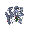
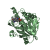
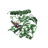
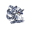
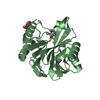
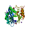
- PDB-7pp0: Crystal structure of the VIM-2 acquired metallo-beta-Lactamase in... -
+ Open data

- Basic information
| Entry | Database: PDB / ID: 7pp0 | ||||||
|---|---|---|---|---|---|---|---|
| Title | Crystal structure of the VIM-2 acquired metallo-beta-Lactamase in complex with compound 28 (JMV-7038) | ||||||
 Components Components | Metallo-beta-lactamase VIM-2-like protein | ||||||
 Keywords Keywords | HYDROLASE / metallo-beta-Lactamase / VIM-2 / triazole-thione / inhibitor / zinc | ||||||
| Function / homology |  Function and homology information Function and homology informationantibiotic catabolic process / beta-lactamase activity / beta-lactamase / periplasmic space / response to antibiotic / metal ion binding Similarity search - Function | ||||||
| Biological species |   Pseudomonas aeruginosa (bacteria) Pseudomonas aeruginosa (bacteria) | ||||||
| Method |  X-RAY DIFFRACTION / SYNCHROTRON / MOLECULAR REPLACEMENT / Resolution: 1.73 Å X-RAY DIFFRACTION / SYNCHROTRON / MOLECULAR REPLACEMENT / Resolution: 1.73 Å | ||||||
 Authors Authors | Tassone, G. / Benvenuti, M. / Verdirosa, F. / Corsica, G. / Chelini, G. / De Luca, F. / Docquier, J.D. / Pozzi, C. / Mangani, S. | ||||||
| Funding support | 1items
| ||||||
 Citation Citation | Journal: Chemmedchem / Year: 2022 Title: 1,2,4-Triazole-3-Thione Analogues with a 2-Ethylbenzoic Acid at Position 4 as VIM-type Metallo-beta-Lactamase Inhibitors. Authors: Verdirosa, F. / Gavara, L. / Sevaille, L. / Tassone, G. / Corsica, G. / Legru, A. / Feller, G. / Chelini, G. / Mercuri, P.S. / Tanfoni, S. / Sannio, F. / Benvenuti, M. / Cerboni, G. / De ...Authors: Verdirosa, F. / Gavara, L. / Sevaille, L. / Tassone, G. / Corsica, G. / Legru, A. / Feller, G. / Chelini, G. / Mercuri, P.S. / Tanfoni, S. / Sannio, F. / Benvenuti, M. / Cerboni, G. / De Luca, F. / Bouajila, E. / Vo Hoang, Y. / Licznar-Fajardo, P. / Galleni, M. / Pozzi, C. / Mangani, S. / Docquier, J.D. / Hernandez, J.F. | ||||||
| History |
|
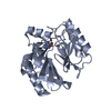
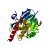
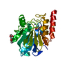
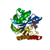
- Structure visualization
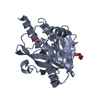
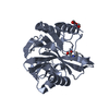
| Structure viewer | Molecule: MolmilJmol/JSmol |
|---|
- Downloads & links
-Download
| PDBx/mmCIF format | 7pp0.cif.gz | 65.6 KB | Display | PDBx/mmCIF format |
|---|---|---|---|---|
| PDB format | pdb7pp0.ent.gz | 45.5 KB | Display | PDB format |
| PDBx/mmJSON format | 7pp0.json.gz | Tree view | PDBx/mmJSON format | |
| Others |  Other downloads Other downloads |
-Validation report
| Arichive directory | https://data.pdbj.org/pub/pdb/validation_reports/pp/7pp0ftp://data.pdbj.org/pub/pdb/validation_reports/pp/7pp0 | HTTPS FTP |
|---|
-Related structure data
| Related structure data |  6sp7S S: Starting model for refinement |
|---|---|
| Similar structure data |


-Links
 PDBj
PDBj

- Assembly
| Deposited unit | 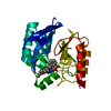
| ||||||||
|---|---|---|---|---|---|---|---|---|---|
| 1 |
| ||||||||
| Unit cell |
| ||||||||
| Components on special symmetry positions |
|
-Components
| #1: Protein | Mass: 25539.322 Da / Num. of mol.: 1 Source method: isolated from a genetically manipulated source Source: (gene. exp.) Pseudomonas aeruginosa (bacteria) / Gene: blaVIM / Plasmid: pET9 / Production host: | ||||||||
|---|---|---|---|---|---|---|---|---|---|
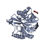
| #2: Chemical |   Mass: 65.409 Da / Num. of mol.: 3 / Source method: obtained synthetically / Formula: Zn Mass: 65.409 Da / Num. of mol.: 3 / Source method: obtained synthetically / Formula: Zn#3: Chemical |   Mass: 59.044 Da / Num. of mol.: 2 / Source method: obtained synthetically / Formula: C2H3O2 Mass: 59.044 Da / Num. of mol.: 2 / Source method: obtained synthetically / Formula: C2H3O2#4: Chemical | ChemComp-7ZN / | 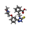  Mass: 454.542 Da / Num. of mol.: 1 / Source method: obtained synthetically / Formula: C23H26N4O4S / Feature type: SUBJECT OF INVESTIGATION Mass: 454.542 Da / Num. of mol.: 1 / Source method: obtained synthetically / Formula: C23H26N4O4S / Feature type: SUBJECT OF INVESTIGATION#5: Water | ChemComp-HOH / |  Mass: 18.015 Da / Num. of mol.: 183 / Source method: isolated from a natural source / Formula: H2O Mass: 18.015 Da / Num. of mol.: 183 / Source method: isolated from a natural source / Formula: H2OHas ligand of interest | Y | |
-Experimental details
-Experiment
| Experiment | Method: X-RAY DIFFRACTION / Number of used crystals: 1 |
|---|
- Sample preparation
| Crystal | Density Matthews: 2.07 Å3/Da / Density % sol: 40.64 % |
|---|---|
| Crystal grow | Temperature: 293 K / Method: vapor diffusion, sitting drop / pH: 6.5 Details: 0.1M cacodilate pH 6.5, 5mM DTT, 0.2M Na Acetate, 26% peg 8000 |
-Data collection
| Diffraction | Mean temperature: 100 K / Serial crystal experiment: N |
|---|---|
| Diffraction source | Source: SYNCHROTRON / Site: Diamond  / Beamline: I04 / Wavelength: 0.9795 Å / Beamline: I04 / Wavelength: 0.9795 Å |
| Detector | Type: DECTRIS EIGER X 16M / Detector: PIXEL / Date: Dec 4, 2019 |
| Radiation | Monochromator: Si(111) / Protocol: SINGLE WAVELENGTH / Monochromatic (M) / Laue (L): M / Scattering type: x-ray |
| Radiation wavelength | Wavelength: 0.9795 Å / Relative weight: 1 |
| Reflection | Resolution: 1.73→55.76 Å / Num. obs: 22531 / % possible obs: 100 % / Observed criterion σ(I): 2 / Redundancy: 7.5 % / Biso Wilson estimate: 18.5 Å2 / CC1/2: 0.999 / Rmerge(I) obs: 0.086 / Rpim(I) all: 0.034 / Rrim(I) all: 0.092 / Net I/σ(I): 12.3 |
| Reflection shell | Resolution: 1.73→1.82 Å / Redundancy: 7.2 % / Rmerge(I) obs: 0.845 / Mean I/σ(I) obs: 2.4 / Num. unique obs: 3242 / CC1/2: 0.821 / Rpim(I) all: 0.337 / Rrim(I) all: 0.911 / % possible all: 100 |
- Processing
| Software |
| |||||||||||||||||||||||||||||||||||||||||||||
|---|---|---|---|---|---|---|---|---|---|---|---|---|---|---|---|---|---|---|---|---|---|---|---|---|---|---|---|---|---|---|---|---|---|---|---|---|---|---|---|---|---|---|---|---|---|---|
| Refinement | Method to determine structure: MOLECULAR REPLACEMENT Starting model: 6SP7 Resolution: 1.73→55.76 Å / Cor.coef. Fo:Fc: 0.968 / Cor.coef. Fo:Fc free: 0.95 / SU B: 2.915 / SU ML: 0.091 / Cross valid method: THROUGHOUT / σ(F): 0 / ESU R: 0.119 / ESU R Free: 0.117 / Stereochemistry target values: MAXIMUM LIKELIHOOD / Details: U VALUES : REFINED INDIVIDUALLY
| |||||||||||||||||||||||||||||||||||||||||||||
| Solvent computation | Ion probe radii: 0.8 Å / Shrinkage radii: 0.8 Å / VDW probe radii: 1.2 Å / Solvent model: MASK | |||||||||||||||||||||||||||||||||||||||||||||
| Displacement parameters | Biso max: 66.73 Å2 / Biso mean: 26.637 Å2 / Biso min: 10.53 Å2
| |||||||||||||||||||||||||||||||||||||||||||||
| Refine analyze | Luzzati coordinate error obs: 0.192 Å | |||||||||||||||||||||||||||||||||||||||||||||
| Refinement step | Cycle: final / Resolution: 1.73→55.76 Å
| |||||||||||||||||||||||||||||||||||||||||||||
| Refine LS restraints |
| |||||||||||||||||||||||||||||||||||||||||||||
| LS refinement shell | Resolution: 1.73→1.775 Å / Rfactor Rfree error: 0 / Total num. of bins used: 20
|
