 Movie
Movie Controller

[English] 日本語
 Yorodumi
- PDB-7mqp: E. coli dihydrofolate reductase complexed with 4'-chloro-3'-(4-(2... -
+ Open data

- Basic information
| Entry | Database: PDB / ID: 7mqp | ||||||
|---|---|---|---|---|---|---|---|
| Title | E. coli dihydrofolate reductase complexed with 4'-chloro-3'-(4-(2,4-diamino-6-ethylpyrimidin-5-yl)but-3-yn-2-yl)-[1,1'-biphenyl]-4-carboxamide (UCP1228) | ||||||
 Components Components | Dihydrofolate reductase | ||||||
 Keywords Keywords | OXIDOREDUCTASE / Inhibitor / antifolate / DHFR | ||||||
| Function / homology |  Function and homology information Function and homology informationNADP+ binding / dihydrofolate metabolic process / dihydrofolate reductase / dihydrofolate reductase activity / folic acid metabolic process / tetrahydrofolate biosynthetic process / one-carbon metabolic process / metal ion binding / cytosol Similarity search - Function | ||||||
| Biological species |  | ||||||
| Method |  X-RAY DIFFRACTION / SYNCHROTRON / MOLECULAR REPLACEMENT / Resolution: 2.1 Å X-RAY DIFFRACTION / SYNCHROTRON / MOLECULAR REPLACEMENT / Resolution: 2.1 Å | ||||||
 Authors Authors | Lombardo, M.N. / Wright, D.L. | ||||||
| Funding support | 1items
| ||||||
 Citation Citation | Journal: Commun Biol / Year: 2022 Title: Structure-guided functional studies of plasmid-encoded dihydrofolate reductases reveal a common mechanism of trimethoprim resistance in Gram-negative pathogens. Authors: Krucinska, J. / Lombardo, M.N. / Erlandsen, H. / Estrada, A. / Si, D. / Viswanathan, K. / Wright, D.L. | ||||||
| History |
|
- Structure visualization
| Structure viewer | Molecule: MolmilJmol/JSmol |
|---|
- Downloads & links
-Download
| PDBx/mmCIF format | 7mqp.cif.gz | 53 KB | Display | PDBx/mmCIF format |
|---|---|---|---|---|
| PDB format | pdb7mqp.ent.gz | 35.7 KB | Display | PDB format |
| PDBx/mmJSON format | 7mqp.json.gz | Tree view | PDBx/mmJSON format | |
| Others |  Other downloads Other downloads |
-Validation report
| Arichive directory | https://data.pdbj.org/pub/pdb/validation_reports/mq/7mqpftp://data.pdbj.org/pub/pdb/validation_reports/mq/7mqp | HTTPS FTP |
|---|
-Related structure data
| Related structure data | 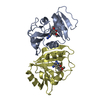 7mylC  7mymC 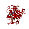 7naeC  7r6gC 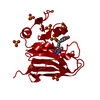 7rebC 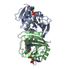 7regC 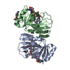 7rgjC 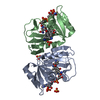 7rgkC 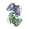 7rgoC 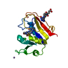 1rf7S S: Starting model for refinement C: citing same article ( |
|---|---|
| Similar structure data |
-Links
 PDBj
PDBj
- Assembly
| Deposited unit | 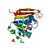
| ||||||||
|---|---|---|---|---|---|---|---|---|---|
| 1 | 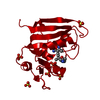
| ||||||||
| Unit cell |
|
-Components
| #1: Protein | Mass: 18262.600 Da / Num. of mol.: 1 Source method: isolated from a genetically manipulated source Source: (gene. exp.) | ||||
|---|---|---|---|---|---|
| #2: Chemical | ChemComp-ZM4 / 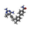  Mass: 399.488 Da / Num. of mol.: 1 / Source method: obtained synthetically / Formula: C24H25N5O / Feature type: SUBJECT OF INVESTIGATION Mass: 399.488 Da / Num. of mol.: 1 / Source method: obtained synthetically / Formula: C24H25N5O / Feature type: SUBJECT OF INVESTIGATION | ||||
| #3: Chemical | ChemComp-SO4 /   Mass: 96.063 Da / Num. of mol.: 7 / Source method: obtained synthetically / Formula: SO4 Mass: 96.063 Da / Num. of mol.: 7 / Source method: obtained synthetically / Formula: SO4#4: Water | ChemComp-HOH / |  Mass: 18.015 Da / Num. of mol.: 57 / Source method: isolated from a natural source / Formula: H2O Mass: 18.015 Da / Num. of mol.: 57 / Source method: isolated from a natural source / Formula: H2OHas ligand of interest | Y | |
-Experimental details
-Experiment
| Experiment | Method: X-RAY DIFFRACTION / Number of used crystals: 1 |
|---|
- Sample preparation
| Crystal | Density Matthews: 3.76 Å3/Da / Density % sol: 67.25 % |
|---|---|
| Crystal grow | Temperature: 277.15 K / Method: vapor diffusion, hanging drop / Details: 0.3-0.35 M lithium sulfate, 15-19% PEG 6,000 |
-Data collection
| Diffraction | Mean temperature: 100 K / Serial crystal experiment: N |
|---|---|
| Diffraction source | Source: SYNCHROTRON / Site: SSRL  / Beamline: BL14-1 / Wavelength: 1 Å / Beamline: BL14-1 / Wavelength: 1 Å |
| Detector | Type: MARMOSAIC 325 mm CCD / Detector: CCD / Date: Nov 16, 2017 |
| Radiation | Protocol: SINGLE WAVELENGTH / Monochromatic (M) / Laue (L): M / Scattering type: x-ray |
| Radiation wavelength | Wavelength: 1 Å / Relative weight: 1 |
| Reflection | Resolution: 2.1→57.81 Å / Num. obs: 17232 / % possible obs: 99.72 % / Redundancy: 1.2 % / CC1/2: 1 / CC star: 1 / Net I/σ(I): 21.82 |
| Reflection shell | Resolution: 2.102→2.177 Å / Redundancy: 1.1 % / Mean I/σ(I) obs: 3.73 / Num. unique obs: 1663 / CC1/2: 1 / CC star: 1 / % possible all: 100 |
- Processing
| Software |
| ||||||||||||||||||||||||||||||||||||||||||||||||||||||||||||
|---|---|---|---|---|---|---|---|---|---|---|---|---|---|---|---|---|---|---|---|---|---|---|---|---|---|---|---|---|---|---|---|---|---|---|---|---|---|---|---|---|---|---|---|---|---|---|---|---|---|---|---|---|---|---|---|---|---|---|---|---|---|
| Refinement | Method to determine structure: MOLECULAR REPLACEMENT Starting model: 1RF7 Resolution: 2.1→57.81 Å / Cor.coef. Fo:Fc: 0.966 / Cor.coef. Fo:Fc free: 0.954 / SU B: 4.225 / SU ML: 0.109 / Cross valid method: THROUGHOUT / σ(F): 0 / ESU R: 0.149 / ESU R Free: 0.147 / Stereochemistry target values: MAXIMUM LIKELIHOOD Details: HYDROGENS HAVE BEEN ADDED IN THE RIDING POSITIONS U VALUES : REFINED INDIVIDUALLY
| ||||||||||||||||||||||||||||||||||||||||||||||||||||||||||||
| Solvent computation | Ion probe radii: 0.8 Å / Shrinkage radii: 0.8 Å / VDW probe radii: 1.2 Å / Solvent model: MASK | ||||||||||||||||||||||||||||||||||||||||||||||||||||||||||||
| Displacement parameters | Biso max: 163.31 Å2 / Biso mean: 65.274 Å2 / Biso min: 42.48 Å2
| ||||||||||||||||||||||||||||||||||||||||||||||||||||||||||||
| Refinement step | Cycle: final / Resolution: 2.1→57.81 Å
| ||||||||||||||||||||||||||||||||||||||||||||||||||||||||||||
| Refine LS restraints |
| ||||||||||||||||||||||||||||||||||||||||||||||||||||||||||||
| LS refinement shell | Resolution: 2.102→2.156 Å / Rfactor Rfree error: 0 / Total num. of bins used: 20
|
