 Movie
Movie Controller

[English] 日本語
 Yorodumi
- PDB-7mym: Crystal structure of Escherichia coli dihydrofolate reductase in ... -
+ Open data

- Basic information
| Entry | Database: PDB / ID: 7mym | ||||||
|---|---|---|---|---|---|---|---|
| Title | Crystal structure of Escherichia coli dihydrofolate reductase in complex with TRIMETHOPRIM and NADPH | ||||||
 Components Components | Dihydrofolate reductase | ||||||
 Keywords Keywords | OXIDOREDUCTASE / dihydrofolate reductase | ||||||
| Function / homology |  Function and homology information Function and homology informationNADP+ binding / dihydrofolate metabolic process / dihydrofolate reductase / dihydrofolate reductase activity / folic acid metabolic process / tetrahydrofolate biosynthetic process / one-carbon metabolic process / cytosol Similarity search - Function | ||||||
| Biological species |  | ||||||
| Method |  X-RAY DIFFRACTION / SYNCHROTRON / MOLECULAR REPLACEMENT / molecular replacement / Resolution: 3.04 Å X-RAY DIFFRACTION / SYNCHROTRON / MOLECULAR REPLACEMENT / molecular replacement / Resolution: 3.04 Å | ||||||
 Authors Authors | Erlandsen, H. / Wright, D. / Krucinska, J. | ||||||
| Funding support |  United States, 1items United States, 1items
| ||||||
 Citation Citation | Journal: Commun Biol / Year: 2022 Title: Structure-guided functional studies of plasmid-encoded dihydrofolate reductases reveal a common mechanism of trimethoprim resistance in Gram-negative pathogens. Authors: Krucinska, J. / Lombardo, M.N. / Erlandsen, H. / Estrada, A. / Si, D. / Viswanathan, K. / Wright, D.L. | ||||||
| History |
|
- Structure visualization
| Structure viewer | Molecule: MolmilJmol/JSmol |
|---|
- Downloads & links
-Download
| PDBx/mmCIF format | 7mym.cif.gz | 117.3 KB | Display | PDBx/mmCIF format |
|---|---|---|---|---|
| PDB format | pdb7mym.ent.gz | 90.2 KB | Display | PDB format |
| PDBx/mmJSON format | 7mym.json.gz | Tree view | PDBx/mmJSON format | |
| Others |  Other downloads Other downloads |
-Validation report
| Arichive directory | https://data.pdbj.org/pub/pdb/validation_reports/my/7mymftp://data.pdbj.org/pub/pdb/validation_reports/my/7mym | HTTPS FTP |
|---|
-Related structure data
| Related structure data | 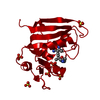 7mqpC 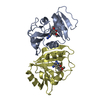 7mylC 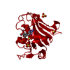 7naeC  7r6gC 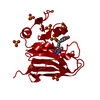 7rebC 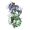 7regC 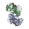 7rgjC 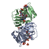 7rgkC 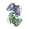 7rgoC  4psyS S: Starting model for refinement C: citing same article ( |
|---|---|
| Similar structure data |
-Links
 PDBj
PDBj

- Assembly
| Deposited unit | 
| ||||||||
|---|---|---|---|---|---|---|---|---|---|
| 1 | 
| ||||||||
| 2 | 
| ||||||||
| 3 | 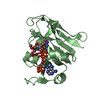
| ||||||||
| Unit cell |
|
-Components
-Protein , 1 types, 3 molecules ACB
| #1: Protein | Mass: 18019.340 Da / Num. of mol.: 3 Source method: isolated from a genetically manipulated source Source: (gene. exp.) |
|---|
-Non-polymers , 5 types, 25 molecules 


| #2: Chemical |  Type: L-peptide linking / Mass: 175.209 Da / Num. of mol.: 2 / Source method: obtained synthetically / Formula: C6H15N4O2 Type: L-peptide linking / Mass: 175.209 Da / Num. of mol.: 2 / Source method: obtained synthetically / Formula: C6H15N4O2#3: Chemical |  Mass: 743.405 Da / Num. of mol.: 3 / Source method: obtained synthetically / Formula: C21H28N7O17P3 / Feature type: SUBJECT OF INVESTIGATION Mass: 743.405 Da / Num. of mol.: 3 / Source method: obtained synthetically / Formula: C21H28N7O17P3 / Feature type: SUBJECT OF INVESTIGATION#4: Chemical |  Mass: 290.318 Da / Num. of mol.: 3 / Source method: obtained synthetically / Formula: C14H18N4O3 / Feature type: SUBJECT OF INVESTIGATION / Comment: antibiotic*YM Mass: 290.318 Da / Num. of mol.: 3 / Source method: obtained synthetically / Formula: C14H18N4O3 / Feature type: SUBJECT OF INVESTIGATION / Comment: antibiotic*YM#5: Chemical | ChemComp-SO4 /  Mass: 96.063 Da / Num. of mol.: 4 / Source method: obtained synthetically / Formula: SO4 Mass: 96.063 Da / Num. of mol.: 4 / Source method: obtained synthetically / Formula: SO4#6: Water | ChemComp-HOH / | Mass: 18.015 Da / Num. of mol.: 13 / Source method: isolated from a natural source / Formula: H2O |
|---|
-Details
| Has ligand of interest | Y |
|---|
-Experimental details
-Experiment
| Experiment | Method: X-RAY DIFFRACTION / Number of used crystals: 1 |
|---|
- Sample preparation
| Crystal | Density Matthews: 3.72 Å3/Da / Density % sol: 66.92 % |
|---|---|
| Crystal grow | Temperature: 277.15 K / Method: vapor diffusion, hanging drop Details: 18% to 20% PEG4000 or 8000, 0.2M ammonium sulfate and 2mM DTT. |
-Data collection
| Diffraction | Mean temperature: 100 K / Serial crystal experiment: N |
|---|---|
| Diffraction source | Source: SYNCHROTRON / Site: NSLS-II / Beamline: 17-ID-1 / Wavelength: 0.9201 Å |
| Detector | Type: DECTRIS EIGER X 9M / Detector: PIXEL / Date: Aug 21, 2020 |
| Radiation | Monochromator: Double Crystal Monochromator, Si111 / Protocol: SINGLE WAVELENGTH / Monochromatic (M) / Laue (L): M / Scattering type: x-ray |
| Radiation wavelength | Wavelength: 0.9201 Å / Relative weight: 1 |
| Reflection | Resolution: 3.04→56.76 Å / Num. obs: 15680 / % possible obs: 98.7 % / Redundancy: 6 % / Biso Wilson estimate: 45.81 Å2 / CC1/2: 0.94 / Rmerge(I) obs: 0.289 / Rpim(I) all: 0.127 / Rrim(I) all: 0.318 / Net I/σ(I): 3.9 |
| Reflection shell | Resolution: 3.04→3.25 Å / Redundancy: 5.8 % / Rmerge(I) obs: 0.863 / Mean I/σ(I) obs: 1.9 / Num. unique obs: 16148 / CC1/2: 0.345 / Rpim(I) all: 0.416 / Rrim(I) all: 0.966 / % possible all: 98.1 |
-Phasing
| Phasing | Method: molecular replacement |
|---|
- Processing
| Software |
| ||||||||||||||||||||||||||||||||||||||||||||||||||||||||||||
|---|---|---|---|---|---|---|---|---|---|---|---|---|---|---|---|---|---|---|---|---|---|---|---|---|---|---|---|---|---|---|---|---|---|---|---|---|---|---|---|---|---|---|---|---|---|---|---|---|---|---|---|---|---|---|---|---|---|---|---|---|---|
| Refinement | Method to determine structure: MOLECULAR REPLACEMENT Starting model: 4PSY Resolution: 3.04→56.76 Å / Cor.coef. Fo:Fc: 0.907 / Cor.coef. Fo:Fc free: 0.822 / SU B: 23.603 / SU ML: 0.406 / Cross valid method: THROUGHOUT / σ(F): 0 / ESU R Free: 0.461 / Stereochemistry target values: MAXIMUM LIKELIHOOD Details: HYDROGENS HAVE BEEN ADDED IN THE RIDING POSITIONS U VALUES : REFINED INDIVIDUALLY
| ||||||||||||||||||||||||||||||||||||||||||||||||||||||||||||
| Solvent computation | Ion probe radii: 0.8 Å / Shrinkage radii: 0.8 Å / VDW probe radii: 1.2 Å / Solvent model: MASK | ||||||||||||||||||||||||||||||||||||||||||||||||||||||||||||
| Displacement parameters | Biso max: 119.68 Å2 / Biso mean: 51.221 Å2 / Biso min: 20.75 Å2
| ||||||||||||||||||||||||||||||||||||||||||||||||||||||||||||
| Refinement step | Cycle: final / Resolution: 3.04→56.76 Å
| ||||||||||||||||||||||||||||||||||||||||||||||||||||||||||||
| Refine LS restraints |
| ||||||||||||||||||||||||||||||||||||||||||||||||||||||||||||
| LS refinement shell | Resolution: 3.04→3.119 Å / Rfactor Rfree error: 0 / Total num. of bins used: 20
|
