 Movie
Movie Controller

[English] 日本語
 Yorodumi
- PDB-7reb: E. coli dihydrofolate reductase complexed with 5-(3-(7-(4-(aminom... -
+ Open data

- Basic information
| Entry | Database: PDB / ID: 7reb | ||||||
|---|---|---|---|---|---|---|---|
| Title | E. coli dihydrofolate reductase complexed with 5-(3-(7-(4-(aminomethyl)phenyl)benzo[d][1,3]dioxol-5-yl)but-1-yn-1-yl)-6-ethylpyrimidine-2,4-diamine (UCP1223) | ||||||
 Components Components | Dihydrofolate reductase | ||||||
 Keywords Keywords | OXIDOREDUCTASE / Inhibitor / antifolate / DHFR | ||||||
| Function / homology |  Function and homology information Function and homology informationNADP+ binding / dihydrofolate metabolic process / dihydrofolate reductase / dihydrofolate reductase activity / folic acid metabolic process / tetrahydrofolate biosynthetic process / one-carbon metabolic process / metal ion binding / cytosol Similarity search - Function | ||||||
| Biological species |  | ||||||
| Method |  X-RAY DIFFRACTION / SYNCHROTRON / MOLECULAR REPLACEMENT / Resolution: 1.91 Å X-RAY DIFFRACTION / SYNCHROTRON / MOLECULAR REPLACEMENT / Resolution: 1.91 Å | ||||||
 Authors Authors | Lombardo, M.N. / Wright, D.L. | ||||||
| Funding support | 1items
| ||||||
 Citation Citation | Journal: Commun Biol / Year: 2022 Title: Structure-guided functional studies of plasmid-encoded dihydrofolate reductases reveal a common mechanism of trimethoprim resistance in Gram-negative pathogens. Authors: Krucinska, J. / Lombardo, M.N. / Erlandsen, H. / Estrada, A. / Si, D. / Viswanathan, K. / Wright, D.L. | ||||||
| History |
|
- Structure visualization
| Structure viewer | Molecule: MolmilJmol/JSmol |
|---|
- Downloads & links
-Download
| PDBx/mmCIF format | 7reb.cif.gz | 59.8 KB | Display | PDBx/mmCIF format |
|---|---|---|---|---|
| PDB format | pdb7reb.ent.gz | 34.7 KB | Display | PDB format |
| PDBx/mmJSON format | 7reb.json.gz | Tree view | PDBx/mmJSON format | |
| Others |  Other downloads Other downloads |
-Validation report
| Arichive directory | https://data.pdbj.org/pub/pdb/validation_reports/re/7rebftp://data.pdbj.org/pub/pdb/validation_reports/re/7reb | HTTPS FTP |
|---|
-Related structure data
| Related structure data |  7mqpC  7mylC  7mymC  7naeC  7r6gC  7regC  7rgjC  7rgkC  7rgoC  1rx2S S: Starting model for refinement C: citing same article ( |
|---|---|
| Similar structure data |
-Links
 PDBj
PDBj
- Assembly
| Deposited unit | 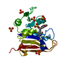
| ||||||||||
|---|---|---|---|---|---|---|---|---|---|---|---|
| 1 | 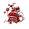
| ||||||||||
| Unit cell |
|
-Components
| #1: Protein | Mass: 18262.600 Da / Num. of mol.: 1 Source method: isolated from a genetically manipulated source Source: (gene. exp.) | ||||||
|---|---|---|---|---|---|---|---|
| #2: Chemical | ChemComp-SO4 /   Mass: 96.063 Da / Num. of mol.: 7 / Source method: obtained synthetically / Formula: SO4 Mass: 96.063 Da / Num. of mol.: 7 / Source method: obtained synthetically / Formula: SO4#3: Chemical | ChemComp-560 / | 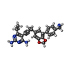  Mass: 415.488 Da / Num. of mol.: 1 / Source method: obtained synthetically / Formula: C24H25N5O2 / Feature type: SUBJECT OF INVESTIGATION Mass: 415.488 Da / Num. of mol.: 1 / Source method: obtained synthetically / Formula: C24H25N5O2 / Feature type: SUBJECT OF INVESTIGATION#4: Water | ChemComp-HOH / |  Mass: 18.015 Da / Num. of mol.: 51 / Source method: isolated from a natural source / Formula: H2O Mass: 18.015 Da / Num. of mol.: 51 / Source method: isolated from a natural source / Formula: H2OHas ligand of interest | Y | |
-Experimental details
-Experiment
| Experiment | Method: X-RAY DIFFRACTION / Number of used crystals: 1 |
|---|
- Sample preparation
| Crystal | Density Matthews: 3.78 Å3/Da / Density % sol: 67.48 % |
|---|---|
| Crystal grow | Temperature: 277 K / Method: vapor diffusion, hanging drop / Details: 0.3-0.35 M lithium sulfate, 15-19% PEG 6,000 |
-Data collection
| Diffraction | Mean temperature: 100 K / Serial crystal experiment: N |
|---|---|
| Diffraction source | Source: SYNCHROTRON / Site: SSRL  / Beamline: BL14-1 / Wavelength: 1 Å / Beamline: BL14-1 / Wavelength: 1 Å |
| Detector | Type: MARMOSAIC 325 mm CCD / Detector: CCD / Date: Jun 15, 2017 |
| Diffraction measurement | Details: 0.25 degrees, 10.85 sec, detector distance 220.00 mm Method: \w scans |
| Radiation | Protocol: SINGLE WAVELENGTH / Monochromatic (M) / Laue (L): M / Scattering type: x-ray |
| Radiation wavelength | Wavelength: 1 Å / Relative weight: 1 |
| Reflection | Av R equivalents: 0.078 / Number: 285948 |
| Reflection | Resolution: 1.91→57.65 Å / Num. obs: 22557 / % possible obs: 97.98 % / Redundancy: 1.2 % / Biso Wilson estimate: 51.19 Å2 / CC1/2: 1 / CC star: 1 / Net I/σ(I): 19.6 |
| Reflection shell | Resolution: 1.91→1.978 Å / Redundancy: 1.1 % / Mean I/σ(I) obs: 2.26 / Num. unique obs: 2128 / CC1/2: 1 / CC star: 1 / % possible all: 96.27 |
| Cell measurement | Reflection used: 285948 |
- Processing
| Software |
| |||||||||||||||||||||||||||||||||||||||||||||||||||||||||||||||
|---|---|---|---|---|---|---|---|---|---|---|---|---|---|---|---|---|---|---|---|---|---|---|---|---|---|---|---|---|---|---|---|---|---|---|---|---|---|---|---|---|---|---|---|---|---|---|---|---|---|---|---|---|---|---|---|---|---|---|---|---|---|---|---|---|
| Refinement | Method to determine structure: MOLECULAR REPLACEMENT Starting model: 1rx2 Resolution: 1.91→34.27 Å / SU ML: 0.4991 / Cross valid method: FREE R-VALUE / σ(F): 1.9 / Phase error: 39.9331 Stereochemistry target values: GeoStd + Monomer Library + CDL v1.2
| |||||||||||||||||||||||||||||||||||||||||||||||||||||||||||||||
| Solvent computation | Shrinkage radii: 0.9 Å / VDW probe radii: 1.11 Å / Solvent model: FLAT BULK SOLVENT MODEL | |||||||||||||||||||||||||||||||||||||||||||||||||||||||||||||||
| Displacement parameters | Biso mean: 67.87 Å2 | |||||||||||||||||||||||||||||||||||||||||||||||||||||||||||||||
| Refinement step | Cycle: LAST / Resolution: 1.91→34.27 Å
| |||||||||||||||||||||||||||||||||||||||||||||||||||||||||||||||
| Refine LS restraints |
| |||||||||||||||||||||||||||||||||||||||||||||||||||||||||||||||
| LS refinement shell |
|
