National Institutes of Health/National Institute of General Medical Sciences (NIH/NIGMS)
R37 GM057247
米国
引用
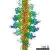
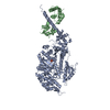
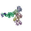
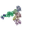
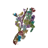
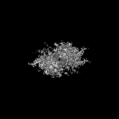
ジャーナル: Proc Natl Acad Sci U S A / 年: 2018 タイトル: High-resolution cryo-EM structures of actin-bound myosin states reveal the mechanism of myosin force sensing. 著者: Ahmet Mentes / Andrew Huehn / Xueqi Liu / Adam Zwolak / Roberto Dominguez / Henry Shuman / E Michael Ostap / Charles V Sindelar / 要旨: Myosins adjust their power outputs in response to mechanical loads in an isoform-dependent manner, resulting in their ability to dynamically adapt to a range of motile challenges. Here, we reveal the ...Myosins adjust their power outputs in response to mechanical loads in an isoform-dependent manner, resulting in their ability to dynamically adapt to a range of motile challenges. Here, we reveal the structural basis for force-sensing based on near-atomic resolution structures of one rigor and two ADP-bound states of myosin-IB (myo1b) bound to actin, determined by cryo-electron microscopy. The two ADP-bound states are separated by a 25° rotation of the lever. The lever of the first ADP state is rotated toward the pointed end of the actin filament and forms a previously unidentified interface with the N-terminal subdomain, which constitutes the upper half of the nucleotide-binding cleft. This pointed-end orientation of the lever blocks ADP release by preventing the N-terminal subdomain from the pivoting required to open the nucleotide binding site, thus revealing how myo1b is inhibited by mechanical loads that restrain lever rotation. The lever of the second ADP state adopts a rigor-like orientation, stabilized by class-specific elements of myo1b. We identify a role for this conformation as an intermediate in the ADP release pathway. Moreover, comparison of our structures with other myosins reveals structural diversity in the actomyosin binding site, and we reveal the high-resolution structure of actin-bound phalloidin, a potent stabilizer of filamentous actin. These results provide a framework to understand the spectrum of force-sensing capacities among the myosin superfamily.
フィルム・検出器のモデル: GATAN K2 SUMMIT (4k x 4k) 平均露光時間: 11.0 sec. / 平均電子線量: 50.0 e/Å2
電子線
加速電圧: 300 kV / 電子線源: FIELD EMISSION GUN
電子光学系
照射モード: SPOT SCAN / 撮影モード: BRIGHT FIELD
実験機器
モデル: Titan Krios / 画像提供: FEI Company
-
画像解析
最終 再構成
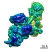
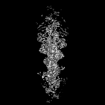
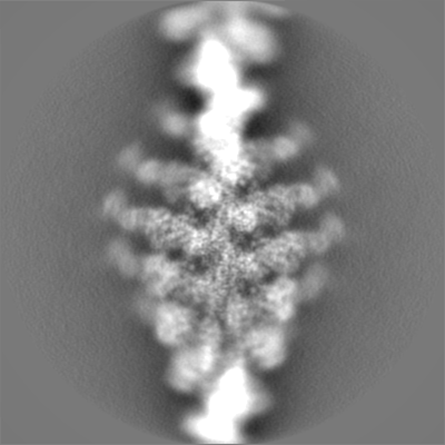
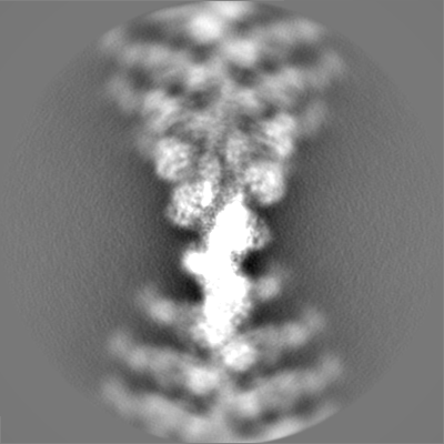
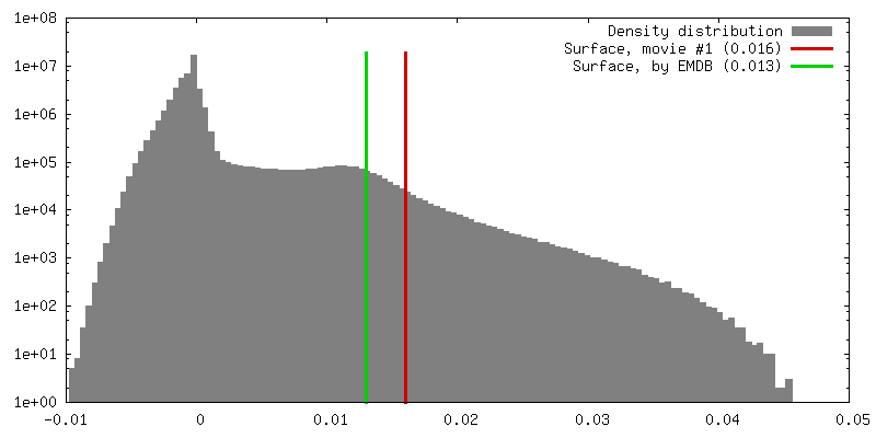
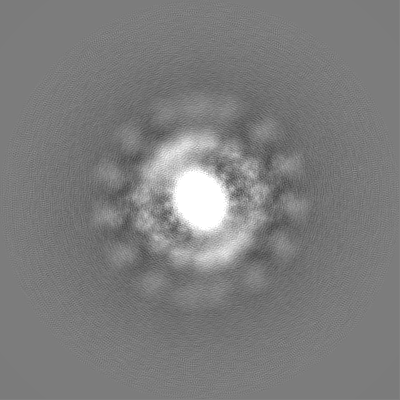
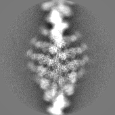
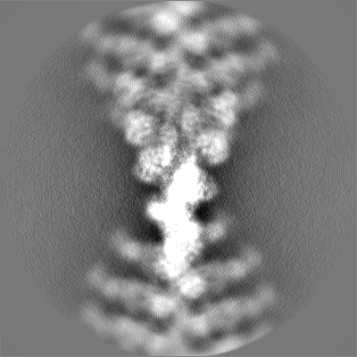
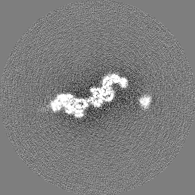
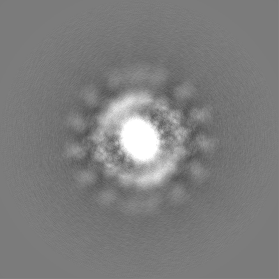
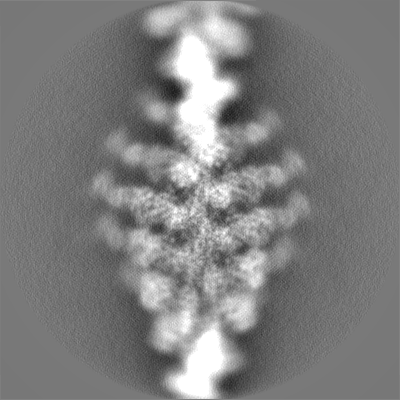
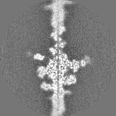
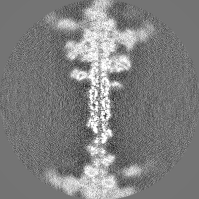
想定した対称性 - らせんパラメータ - Δz: 27.5 Å 想定した対称性 - らせんパラメータ - ΔΦ: -167.4 ° 想定した対称性 - らせんパラメータ - 軸対称性: C1 (非対称) 解像度のタイプ: BY AUTHOR / 解像度: 3.9 Å / 解像度の算出法: FSC 0.143 CUT-OFF 詳細: Resolution estimated by post-processing in RELION using a mask with soft edges that included only the central subunit. 使用した粒子像数: 62000
CTF補正
タイプ: PHASE FLIPPING AND AMPLITUDE CORRECTION
初期モデル
モデルのタイプ: OTHER
最終 角度割当
タイプ: NOT APPLICABLE
-
原子モデル構築 1
精密化
空間: REAL / プロトコル: FLEXIBLE FIT
得られたモデル
PDB-6c1h: High-Resolution Cryo-EM Structures of Actin-bound Myosin States Reveal the Mechanism of Myosin Force Sensing
 ムービー
ムービー コントローラー


 データを開く
 基本情報
基本情報 マップデータ
 試料
試料 キーワード
キーワード 機能・相同性情報
 データ登録者
 米国, 1件
  引用
引用 構造の表示
構造の表示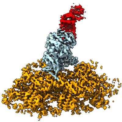
 ダウンロードとリンク
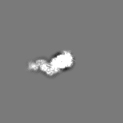
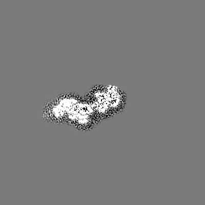
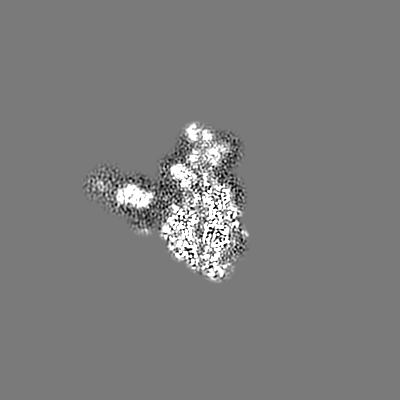
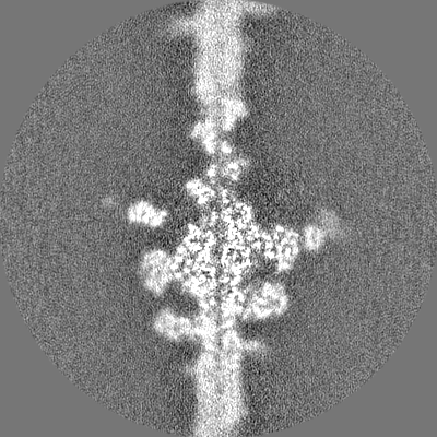
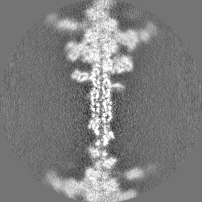
 emd_7331.png
 http://ftp.pdbj.org/pub/emdb/structures/EMD-7331
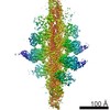


 Z (Sec.)
 Y (Row.)
 X (Col.)


 試料の構成要素
 解析
解析 電子顕微鏡法
 FIELD EMISSION GUN

