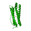
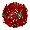
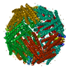
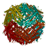
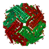
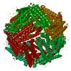
ジャーナル: J Am Chem Soc / 年: 2020 タイトル: Folding of an Intrinsically Disordered Iron-Binding Peptide in Response to Sedimentation Revealed by Cryo-EM. 著者: Geula Davidov / Gili Abelya / Ran Zalk / Benjamin Izbicki / Sharon Shaibi / Lior Spektor / Dayana Shagidov / Esther G Meyron-Holtz / Raz Zarivach / Gabriel A Frank / 要旨: Biomineralization is mediated by specialized proteins that guide and control mineral sedimentation. In many cases, the active regions of these biomineralization proteins are intrinsically disordered. ...Biomineralization is mediated by specialized proteins that guide and control mineral sedimentation. In many cases, the active regions of these biomineralization proteins are intrinsically disordered. High-resolution structures of these proteins while they interact with minerals are essential for understanding biomineralization processes and the function of intrinsically disordered proteins (IDPs). Here we used the cavity of ferritin as a nanoreactor where the interaction between M6A, an intrinsically disordered iron-binding domain, and an iron oxide particle was visualized at high resolution by cryo-EM. Taking advantage of the differences in the electron-dose sensitivity of the protein and the iron oxide particles, we developed a method to determine the irregular shape of the particles found in our density maps. We found that the folding of M6A correlates with the detection of mineral particles in its vicinity. M6A interacts with the iron oxide particles through its C-terminal side, resulting in the stabilization of a helix at its N-terminal side. The stabilization of the helix at a region that is not in direct contact with the iron oxide particle demonstrates the ability of IDPs to respond to signals from their surroundings by conformational changes. These findings provide the first glimpse toward the long-suspected mechanism for biomineralization protein control over mineral microstructure, where unstructured regions of these proteins become more ordered in response to their interaction with the nascent mineral particles.
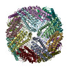
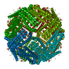
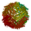
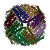
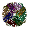
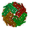
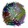
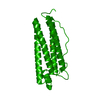
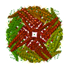
名称: Iron-loaded L-Ferritin-M6A / タイプ: COMPLEX 詳細: Nano cage L_ferritin_M6A at 0.1 mg per mL concentration after sodium acetate treatment with 0.044 mM FeCl2, Iron loaded Entity ID: all / 由来: RECOMBINANT
 ムービー
ムービー コントローラー


 データを開く
 基本情報
基本情報 要素
要素 フェリチン
フェリチン  キーワード
キーワード 機能・相同性情報
 データ登録者
 イスラエル, 2件
  引用
引用 構造の表示
構造の表示 ダウンロードとリンク
 その他のダウンロード
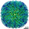


 PDBj
PDBj


 集合体
集合体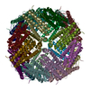

 試料調製
試料調製 電子顕微鏡撮影
 解析
解析