 Movie
Movie Controller

[English] 日本語
 Yorodumi
- EMDB-11265: Folding of an iron binding peptide in response to sedimentation i... -
+ Open data

- Basic information
| Entry | Database: EMDB / ID: EMD-11265 | |||||||||
|---|---|---|---|---|---|---|---|---|---|---|
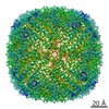
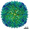
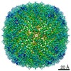
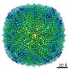
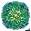
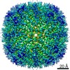
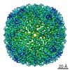
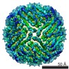
| Title | Folding of an iron binding peptide in response to sedimentation is resolved using ferritin as a nano-reactor | |||||||||
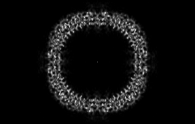
 Map data Map data | Acetate treated L-Ferritin-M6A fusion protein. | |||||||||
 Sample Sample |
| |||||||||
 Keywords Keywords | Biomineralization / nano-reactor / radiation damage assisted single-particle analysis / METAL BINDING PROTEIN | |||||||||
| Function / homology |  Function and homology information Function and homology informationautolysosome / ferric iron binding / iron ion transport / cytoplasmic vesicle / intracellular iron ion homeostasis Similarity search - Function | |||||||||
| Biological species |  | |||||||||
| Method | single particle reconstruction / cryo EM / Resolution: 3.0 Å | |||||||||
 Authors Authors | Davidov G / Abelya G | |||||||||
| Funding support |  Israel, 2 items Israel, 2 items
| |||||||||
 Citation Citation | Journal: J Am Chem Soc / Year: 2020 Title: Folding of an Intrinsically Disordered Iron-Binding Peptide in Response to Sedimentation Revealed by Cryo-EM. Authors: Geula Davidov / Gili Abelya / Ran Zalk / Benjamin Izbicki / Sharon Shaibi / Lior Spektor / Dayana Shagidov / Esther G Meyron-Holtz / Raz Zarivach / Gabriel A Frank / Abstract: Biomineralization is mediated by specialized proteins that guide and control mineral sedimentation. In many cases, the active regions of these biomineralization proteins are intrinsically disordered. ...Biomineralization is mediated by specialized proteins that guide and control mineral sedimentation. In many cases, the active regions of these biomineralization proteins are intrinsically disordered. High-resolution structures of these proteins while they interact with minerals are essential for understanding biomineralization processes and the function of intrinsically disordered proteins (IDPs). Here we used the cavity of ferritin as a nanoreactor where the interaction between M6A, an intrinsically disordered iron-binding domain, and an iron oxide particle was visualized at high resolution by cryo-EM. Taking advantage of the differences in the electron-dose sensitivity of the protein and the iron oxide particles, we developed a method to determine the irregular shape of the particles found in our density maps. We found that the folding of M6A correlates with the detection of mineral particles in its vicinity. M6A interacts with the iron oxide particles through its C-terminal side, resulting in the stabilization of a helix at its N-terminal side. The stabilization of the helix at a region that is not in direct contact with the iron oxide particle demonstrates the ability of IDPs to respond to signals from their surroundings by conformational changes. These findings provide the first glimpse toward the long-suspected mechanism for biomineralization protein control over mineral microstructure, where unstructured regions of these proteins become more ordered in response to their interaction with the nascent mineral particles. | |||||||||
| History |
|
- Structure visualization
| Movie |
Movie viewer |
|---|---|
| Structure viewer | EM map: SurfViewMolmilJmol/JSmol |
| Supplemental images |
- Downloads & links
-EMDB archive
| Map data | emd_11265.map.gz | 20.7 MB | EMDB map data format | |
|---|---|---|---|---|
| Header (meta data) | emd-11265-v30.xmlemd-11265.xml | 14.2 KB 14.2 KB | Display Display | EMDB header |
| Images |  emd_11265.png emd_11265.png | 68.5 KB | ||
| Filedesc metadata | emd-11265.cif.gz | 5.8 KB | ||
| Archive directory |  http://ftp.pdbj.org/pub/emdb/structures/EMD-11265ftp://ftp.pdbj.org/pub/emdb/structures/EMD-11265 http://ftp.pdbj.org/pub/emdb/structures/EMD-11265ftp://ftp.pdbj.org/pub/emdb/structures/EMD-11265 | HTTPS FTP |
-Related structure data
| Related structure data |  6zlgMC  6z3dC  6zh5C  6zlqC C: citing same article ( M: atomic model generated by this map |
|---|---|
| Similar structure data |


-Links
| EMDB pages | EMDB (EBI/PDBe) / EMDataResource |
|---|---|
| Related items in Molecule of the Month |


-Map
| File | Download / File: emd_11265.map.gz / Format: CCP4 / Size: 64 MB / Type: IMAGE STORED AS FLOATING POINT NUMBER (4 BYTES) | ||||||||||||||||||||||||||||||||||||||||||||||||||||||||||||
|---|---|---|---|---|---|---|---|---|---|---|---|---|---|---|---|---|---|---|---|---|---|---|---|---|---|---|---|---|---|---|---|---|---|---|---|---|---|---|---|---|---|---|---|---|---|---|---|---|---|---|---|---|---|---|---|---|---|---|---|---|---|
| Annotation | Acetate treated L-Ferritin-M6A fusion protein. | ||||||||||||||||||||||||||||||||||||||||||||||||||||||||||||
| Projections & slices | Image control
Images are generated by Spider. | ||||||||||||||||||||||||||||||||||||||||||||||||||||||||||||
| Voxel size | X=Y=Z: 1.1 Å | ||||||||||||||||||||||||||||||||||||||||||||||||||||||||||||
| Density |
| ||||||||||||||||||||||||||||||||||||||||||||||||||||||||||||
| Symmetry | Space group: 1 | ||||||||||||||||||||||||||||||||||||||||||||||||||||||||||||
| Details | EMDB XML:
CCP4 map header:
| ||||||||||||||||||||||||||||||||||||||||||||||||||||||||||||
 Z (Sec.)
 Y (Row.)
 X (Col.)


-Supplemental data
- Sample components
-Entire : Iron-loaded L-Ferritin-M6A
| Entire | Name: Iron-loaded L-Ferritin-M6A |
|---|---|
| Components |
|
-Supramolecule #1: Iron-loaded L-Ferritin-M6A
| Supramolecule | Name: Iron-loaded L-Ferritin-M6A / type: complex / ID: 1 / Parent: 0 / Macromolecule list: all Details: Nano cage L_ferritin_M6A at 0.1 mg per mL concentration after sodium acetate treatment with 0.044 mM FeCl2, Iron loaded |
|---|---|
| Source (natural) | Organism: |
-Macromolecule #1: Ferritin
| Macromolecule | Name: Ferritin / type: protein_or_peptide / ID: 1 / Number of copies: 24 / Enantiomer: LEVO |
|---|---|
| Source (natural) | Organism: |
| Molecular weight | Theoretical: 24.362076 KDa |
| Recombinant expression | Organism:  |
| Sequence | String: MGSSHHHHHH SSGLVPRGSH MTSQIRQNYS TEVEAAVNRL VNLHLRASYT YLSLGFFFDR DDVALEGVGH FFRELAEEKR EGAERLLEF QNDRGGRALF QDVQKPSQDE WGKTQEAMEA ALAMEKNLNQ ALLDLHALGS ARADPHLCDF LESHYLDKEV K LIKKMGNH ...String: MGSSHHHHHH SSGLVPRGSH MTSQIRQNYS TEVEAAVNRL VNLHLRASYT YLSLGFFFDR DDVALEGVGH FFRELAEEKR EGAERLLEF QNDRGGRALF QDVQKPSQDE WGKTQEAMEA ALAMEKNLNQ ALLDLHALGS ARADPHLCDF LESHYLDKEV K LIKKMGNH LTNLRRVAGP QPAQTGAPQG SLGEYLFERL TLKHDGDIES AQSDEEVE UniProtKB: Ferritin |
-Experimental details
-Structure determination
| Method | cryo EM |
|---|---|
 Processing Processing | single particle reconstruction |
| Aggregation state | particle |
-Sample preparation
| Concentration | 0.1 mg/mL |
|---|---|
| Buffer | pH: 5.8 |
| Vitrification | Cryogen name: ETHANE |
- Electron microscopy
| Microscope | FEI POLARA 300 |
|---|---|
| Image recording | Film or detector model: GATAN K2 SUMMIT (4k x 4k) / Detector mode: COUNTING / Average electron dose: 80.0 e/Å2 |
| Electron beam | Acceleration voltage: 300 kV / Electron source:  FIELD EMISSION GUN FIELD EMISSION GUN |
| Electron optics | Illumination mode: FLOOD BEAM / Imaging mode: BRIGHT FIELD |
| Experimental equipment |  Model: Tecnai Polara / Image courtesy: FEI Company |
