Movie
Movie Controller


+ Open data

- Basic information
| Entry | Database: PDB / ID: 6zgj | |||||||||
|---|---|---|---|---|---|---|---|---|---|---|
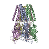
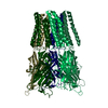
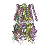
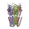
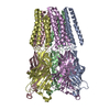
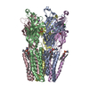
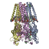
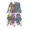
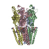
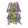
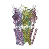
| Title | GLIC pentameric ligand-gated ion channel, pH 5 | |||||||||
 Components Components | Proton-gated ion channel | |||||||||
 Keywords Keywords | MEMBRANE PROTEIN / GLIC / pentameric ligand-gated ion channel / cryoem | |||||||||
| Function / homology |  Function and homology information Function and homology informationsodium channel activity / extracellular ligand-gated monoatomic ion channel activity / potassium channel activity / transmembrane transporter complex / regulation of membrane potential / transmembrane signaling receptor activity / neuron projection / signal transduction / identical protein binding / plasma membrane Similarity search - Function | |||||||||
| Biological species |  Gloeobacter violaceus (bacteria) Gloeobacter violaceus (bacteria) | |||||||||
| Method | ELECTRON MICROSCOPY / single particle reconstruction / cryo EM / Resolution: 3.4 Å | |||||||||
 Authors Authors | Rovsnik, U. / Zhuang, Y. / Forsberg, B.O. / Carroni, M. / Yvonnesdotter, L. / Howard, R.J. / Lindahl, E. | |||||||||
| Funding support |  Sweden, 2items Sweden, 2items
| |||||||||
 Citation Citation | Journal: Life Sci Alliance / Year: 2021 Title: Dynamic closed states of a ligand-gated ion channel captured by cryo-EM and simulations. Authors: Urška Rovšnik / Yuxuan Zhuang / Björn O Forsberg / Marta Carroni / Linnea Yvonnesdotter / Rebecca J Howard / Erik Lindahl /  Abstract: Ligand-gated ion channels are critical mediators of electrochemical signal transduction across evolution. Biophysical and pharmacological characterization of these receptor proteins relies on high- ...Ligand-gated ion channels are critical mediators of electrochemical signal transduction across evolution. Biophysical and pharmacological characterization of these receptor proteins relies on high-quality structures in multiple, subtly distinct functional states. However, structural data in this family remain limited, particularly for resting and intermediate states on the activation pathway. Here, we report cryo-electron microscopy (cryo-EM) structures of the proton-activated ligand-gated ion channel (GLIC) under three pH conditions. Decreased pH was associated with improved resolution and side chain rearrangements at the subunit/domain interface, particularly involving functionally important residues in the β1-β2 and M2-M3 loops. Molecular dynamics simulations substantiated flexibility in the closed-channel extracellular domains relative to the transmembrane ones and supported electrostatic remodeling around E35 and E243 in proton-induced gating. Exploration of secondary cryo-EM classes further indicated a low-pH population with an expanded pore. These results allow us to define distinct protonation and activation steps in pH-stimulated conformational cycling in GLIC, including interfacial rearrangements largely conserved in the pentameric channel family. | |||||||||
| History |
|
- Structure visualization
| Movie |
Movie viewer |
|---|---|
| Structure viewer | Molecule: MolmilJmol/JSmol |
- Downloads & links
-Download
| PDBx/mmCIF format | 6zgj.cif.gz | 262.4 KB | Display | PDBx/mmCIF format |
|---|---|---|---|---|
| PDB format | pdb6zgj.ent.gz | 209.1 KB | Display | PDB format |
| PDBx/mmJSON format | 6zgj.json.gz | Tree view | PDBx/mmJSON format | |
| Others |  Other downloads Other downloads |
-Validation report
| Summary document | 6zgj_validation.pdf.gz | 1 MB | Display | wwPDB validaton report |
|---|---|---|---|---|
| Full document | 6zgj_full_validation.pdf.gz | 1 MB | Display | |
| Data in XML | 6zgj_validation.xml.gz | 54.1 KB | Display | |
| Data in CIF | 6zgj_validation.cif.gz | 75.2 KB | Display | |
| Arichive directory | https://data.pdbj.org/pub/pdb/validation_reports/zg/6zgjftp://data.pdbj.org/pub/pdb/validation_reports/zg/6zgj | HTTPS FTP |
-Related structure data
| Related structure data |  11208MC  6zgdC  6zgkC M: map data used to model this data C: citing same article ( |
|---|---|
| Similar structure data |


-Links
 PDBj
PDBj

- Assembly
| Deposited unit | 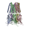
|
|---|---|
| 1 |
|
-Components
| #1: Protein | Mass: 36291.750 Da / Num. of mol.: 5 Source method: isolated from a genetically manipulated source Source: (gene. exp.) Gloeobacter violaceus (strain ATCC 29082 / PCC 7421) (bacteria)Strain: ATCC 29082 / PCC 7421 / Gene: glvI, glr4197 / Production host: |
|---|
-Experimental details
-Experiment
| Experiment | Method: ELECTRON MICROSCOPY |
|---|---|
| EM experiment | Aggregation state: PARTICLE / 3D reconstruction method: single particle reconstruction |
- Sample preparation
| Component | Name: Pentameric ion channel / Type: COMPLEX / Entity ID: all / Source: RECOMBINANT |
|---|---|
| Molecular weight | Value: 0.18 MDa / Experimental value: NO |
| Source (natural) | Organism: Gloeobacter violaceus (strain ATCC 29082 / PCC 7421) (bacteria) |
| Source (recombinant) | Organism: |
| Buffer solution | pH: 7.4 |
| Specimen | Conc.: 3 mg/ml / Embedding applied: NO / Shadowing applied: NO / Staining applied: NO / Vitrification applied: YES |
| Vitrification | Instrument: FEI VITROBOT MARK IV / Cryogen name: ETHANE / Humidity: 100 % / Chamber temperature: 295 K / Details: blot for 1.5 s force -1 |
- Electron microscopy imaging
| Experimental equipment |  Model: Titan Krios / Image courtesy: FEI Company |
|---|---|
| Microscopy | Model: FEI TITAN KRIOS |
| Electron gun | Electron source:  FIELD EMISSION GUN / Accelerating voltage: 300 kV / Illumination mode: FLOOD BEAM FIELD EMISSION GUN / Accelerating voltage: 300 kV / Illumination mode: FLOOD BEAM |
| Electron lens | Mode: BRIGHT FIELD / Nominal magnification: 165000 X / Nominal defocus max: 3800 nm / Nominal defocus min: 2000 nm / Calibrated defocus min: 0.9 nm / Calibrated defocus max: 2.8 nm / Cs: 2.7 mm / C2 aperture diameter: 70 µm / Alignment procedure: COMA FREE |
| Specimen holder | Cryogen: NITROGEN / Specimen holder model: FEI TITAN KRIOS AUTOGRID HOLDER / Temperature (min): 80 K |
| Image recording | Average exposure time: 6 sec. / Electron dose: 45 e/Å2 / Detector mode: COUNTING / Film or detector model: GATAN K2 SUMMIT (4k x 4k) / Num. of grids imaged: 1 / Num. of real images: 6000 |
| EM imaging optics | Energyfilter name: GIF Bioquantum / Energyfilter slit width: 20 eV |
| Image scans | Movie frames/image: 40 / Used frames/image: 1-40 |
- Processing
| Software | Name: PHENIX / Version: 1.18.2_3874: / Classification: refinement | ||||||||||||||||||||||||||||
|---|---|---|---|---|---|---|---|---|---|---|---|---|---|---|---|---|---|---|---|---|---|---|---|---|---|---|---|---|---|
| EM software |
| ||||||||||||||||||||||||||||
| CTF correction | Type: PHASE FLIPPING AND AMPLITUDE CORRECTION | ||||||||||||||||||||||||||||
| Particle selection | Num. of particles selected: 700000 | ||||||||||||||||||||||||||||
| 3D reconstruction | Resolution: 3.4 Å / Resolution method: FSC 0.143 CUT-OFF / Num. of particles: 300000 / Symmetry type: POINT | ||||||||||||||||||||||||||||
| Atomic model building | B value: 278 / Protocol: FLEXIBLE FIT / Space: REAL | ||||||||||||||||||||||||||||
| Atomic model building | PDB-ID: 4NPQ Pdb chain-ID: A / Accession code: 4NPQ / Pdb chain residue range: 5-315 / Source name: PDB / Type: experimental model | ||||||||||||||||||||||||||||
| Refine LS restraints |
|