Movie
Movie Controller


+ Open data

- Basic information
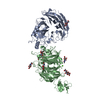
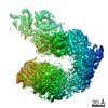
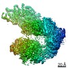
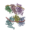
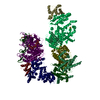
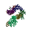
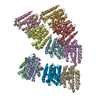
| Entry | Database: PDB / ID: 6vxk | ||||||||||||||||||
|---|---|---|---|---|---|---|---|---|---|---|---|---|---|---|---|---|---|---|---|
| Title | Cryo-EM Structure of the full-length A39R/PlexinC1 complex | ||||||||||||||||||
 Components Components |
| ||||||||||||||||||
 Keywords Keywords | MEMBRANE PROTEIN / receptor / signaling / plexin | ||||||||||||||||||
| Function / homology |  Function and homology information Function and homology informationsemaphorin receptor binding / Other semaphorin interactions / cerebellar climbing fiber to Purkinje cell synapse / regulation of synapse pruning / semaphorin receptor complex / chemorepellent activity / semaphorin receptor activity / negative regulation of cell adhesion / positive regulation of axonogenesis / semaphorin-plexin signaling pathway ...semaphorin receptor binding / Other semaphorin interactions / cerebellar climbing fiber to Purkinje cell synapse / regulation of synapse pruning / semaphorin receptor complex / chemorepellent activity / semaphorin receptor activity / negative regulation of cell adhesion / positive regulation of axonogenesis / semaphorin-plexin signaling pathway / synapse assembly / regulation of cell migration / integrin-mediated signaling pathway / integrin binding / regulation of cell shape / cell adhesion / positive regulation of cell migration / signaling receptor binding / extracellular region / membrane / plasma membrane Similarity search - Function | ||||||||||||||||||
| Biological species |  Ectromelia virus Ectromelia virus Homo sapiens (human) Homo sapiens (human) | ||||||||||||||||||
| Method | ELECTRON MICROSCOPY / single particle reconstruction / cryo EM / Resolution: 3.1 Å | ||||||||||||||||||
 Authors Authors | Kuo, Y.-C. / Chen, H. / Shang, G. / Uchikawa, E. / Tian, H. / Bai, X. / Zhang, X. | ||||||||||||||||||
| Funding support |  United States, 5items United States, 5items
| ||||||||||||||||||
 Citation Citation | Journal: Nat Commun / Year: 2020 Title: Cryo-EM structure of the PlexinC1/A39R complex reveals inter-domain interactions critical for ligand-induced activation. Authors: Yi-Chun Kuo / Hua Chen / Guijun Shang / Emiko Uchikawa / Hui Tian / Xiao-Chen Bai / Xuewu Zhang / Abstract: Plexins are receptors for semaphorins that transduce signals for regulating neuronal development and other processes. Plexins are single-pass transmembrane proteins with multiple domains in both the ...Plexins are receptors for semaphorins that transduce signals for regulating neuronal development and other processes. Plexins are single-pass transmembrane proteins with multiple domains in both the extracellular and intracellular regions. Semaphorin activates plexin by binding to its extracellular N-terminal Sema domain, inducing the active dimer of the plexin intracellular region. The mechanism underlying this activation process of plexin is incompletely understood. We present cryo-electron microscopic structure of full-length human PlexinC1 in complex with the viral semaphorin mimic A39R. The structure shows that A39R induces a specific dimer of PlexinC1 where the membrane-proximal domains from the two PlexinC1 protomers are placed close to each other, poised to promote the active dimer of the intracellular region. This configuration is imposed by a distinct conformation of the PlexinC1 extracellular region, stabilized by inter-domain interactions among the Sema and membrane-proximal domains. Our mutational analyses support the critical role of this conformation in PlexinC1 activation. | ||||||||||||||||||
| History |
|
- Structure visualization
| Movie |
Movie viewer |
|---|---|
| Structure viewer | Molecule: MolmilJmol/JSmol |
- Downloads & links
-Download
| PDBx/mmCIF format | 6vxk.cif.gz | 442.6 KB | Display | PDBx/mmCIF format |
|---|---|---|---|---|
| PDB format | pdb6vxk.ent.gz | 324.5 KB | Display | PDB format |
| PDBx/mmJSON format | 6vxk.json.gz | Tree view | PDBx/mmJSON format | |
| Others |  Other downloads Other downloads |
-Validation report
| Arichive directory | https://data.pdbj.org/pub/pdb/validation_reports/vx/6vxkftp://data.pdbj.org/pub/pdb/validation_reports/vx/6vxk | HTTPS FTP |
|---|
-Related structure data
| Related structure data | 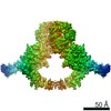 21442MC M: map data used to model this data C: citing same article ( |
|---|---|
| Similar structure data |
-Links
 PDBj
PDBj


- Assembly
| Deposited unit | 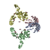
|
|---|---|
| 1 |
|
-Components
| #1: Protein | Mass: 45330.195 Da / Num. of mol.: 2 Source method: isolated from a genetically manipulated source Source: (gene. exp.) Ectromelia virus (strain Moscow) / Strain: Moscow / Gene: EVM139, SEMA / Production host: Homo sapiens (human) / References: UniProt: Q8JL80#2: Protein | Mass: 173487.000 Da / Num. of mol.: 2 Source method: isolated from a genetically manipulated source Source: (gene. exp.) Homo sapiens (human) / Gene: PLXNC1, VESPR / Production host: Homo sapiens (human) / References: UniProt: O60486#3: Sugar | ChemComp-NAG /   Type: D-saccharide, beta linking / Mass: 221.208 Da / Num. of mol.: 18 Type: D-saccharide, beta linking / Mass: 221.208 Da / Num. of mol.: 18Source method: isolated from a genetically manipulated source Formula: C8H15NO6 Has ligand of interest | N | Has protein modification | Y | |
|---|
-Experimental details
-Experiment
| Experiment | Method: ELECTRON MICROSCOPY |
|---|---|
| EM experiment | Aggregation state: PARTICLE / 3D reconstruction method: single particle reconstruction |
- Sample preparation
| Component |
| ||||||||||||||||||||||||||||
|---|---|---|---|---|---|---|---|---|---|---|---|---|---|---|---|---|---|---|---|---|---|---|---|---|---|---|---|---|---|
| Molecular weight |
| ||||||||||||||||||||||||||||
| Source (natural) |
| ||||||||||||||||||||||||||||
| Source (recombinant) |
| ||||||||||||||||||||||||||||
| Buffer solution | pH: 7.5 | ||||||||||||||||||||||||||||
| Buffer component |
| ||||||||||||||||||||||||||||
| Specimen | Conc.: 0.5 mg/ml / Embedding applied: NO / Shadowing applied: NO / Staining applied: NO / Vitrification applied: YES | ||||||||||||||||||||||||||||
| Specimen support | Grid type: Quantifoil, UltrAuFoil, R1.2/1.3 | ||||||||||||||||||||||||||||
| Vitrification | Cryogen name: NITROGEN |
- Electron microscopy imaging
| Experimental equipment |  Model: Titan Krios / Image courtesy: FEI Company |
|---|---|
| Microscopy | Model: FEI TITAN KRIOS |
| Electron gun | Electron source:  FIELD EMISSION GUN / Accelerating voltage: 300 kV / Illumination mode: FLOOD BEAM FIELD EMISSION GUN / Accelerating voltage: 300 kV / Illumination mode: FLOOD BEAM |
| Electron lens | Mode: BRIGHT FIELD |
| Image recording | Electron dose: 2 e/Å2 / Film or detector model: GATAN K3 BIOQUANTUM (6k x 4k) |
- Processing
| Software |
| ||||||||||||||||||||||||
|---|---|---|---|---|---|---|---|---|---|---|---|---|---|---|---|---|---|---|---|---|---|---|---|---|---|
| CTF correction | Type: PHASE FLIPPING AND AMPLITUDE CORRECTION | ||||||||||||||||||||||||
| Symmetry | Point symmetry: C2 (2 fold cyclic) | ||||||||||||||||||||||||
| 3D reconstruction | Resolution: 3.1 Å / Resolution method: FSC 0.143 CUT-OFF / Num. of particles: 143750 / Symmetry type: POINT | ||||||||||||||||||||||||
| Atomic model building | Protocol: AB INITIO MODEL / Space: REAL | ||||||||||||||||||||||||
| Atomic model building | PDB-ID: 3NVN Accession code: 3NVN / Source name: PDB / Type: experimental model | ||||||||||||||||||||||||
| Refinement | Cross valid method: NONE Stereochemistry target values: GeoStd + Monomer Library + CDL v1.2 | ||||||||||||||||||||||||
| Displacement parameters | Biso mean: 100.65 Å2 | ||||||||||||||||||||||||
| Refine LS restraints |
|