Movie
Movie Controller


+ Open data

- Basic information
| Entry | Database: EMDB / ID: EMD-21442 | ||||||||||||||||||
|---|---|---|---|---|---|---|---|---|---|---|---|---|---|---|---|---|---|---|---|
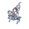
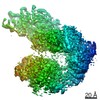
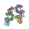
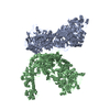
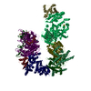
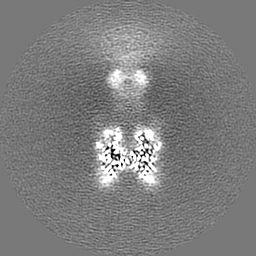
| Title | Cryo-EM Structure of the full-length A39R/PlexinC1 complex | ||||||||||||||||||
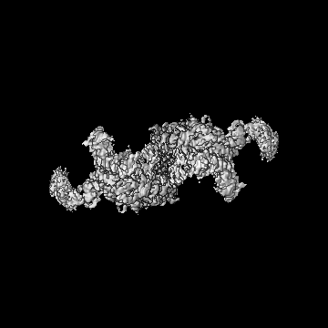
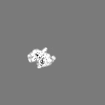
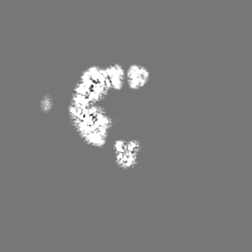
 Map data Map data | Dimeric map | ||||||||||||||||||
 Sample Sample |
| ||||||||||||||||||
 Keywords Keywords | receptor / signaling / plexin / MEMBRANE PROTEIN | ||||||||||||||||||
| Function / homology |  Function and homology information Function and homology informationsemaphorin receptor binding / Other semaphorin interactions / cerebellar climbing fiber to Purkinje cell synapse / regulation of synapse pruning / semaphorin receptor complex / chemorepellent activity / semaphorin receptor activity / negative regulation of cell adhesion / positive regulation of axonogenesis / semaphorin-plexin signaling pathway ...semaphorin receptor binding / Other semaphorin interactions / cerebellar climbing fiber to Purkinje cell synapse / regulation of synapse pruning / semaphorin receptor complex / chemorepellent activity / semaphorin receptor activity / negative regulation of cell adhesion / positive regulation of axonogenesis / semaphorin-plexin signaling pathway / synapse assembly / regulation of cell migration / integrin-mediated signaling pathway / integrin binding / regulation of cell shape / cell adhesion / positive regulation of cell migration / signaling receptor binding / extracellular region / membrane / plasma membrane Similarity search - Function | ||||||||||||||||||
| Biological species |  Homo sapiens (human) / Homo sapiens (human) /  Ectromelia virus / Ectromelia virus (strain Moscow) Ectromelia virus / Ectromelia virus (strain Moscow) | ||||||||||||||||||
| Method | single particle reconstruction / cryo EM / Resolution: 3.1 Å | ||||||||||||||||||
 Authors Authors | Kuo Y-C / Chen H | ||||||||||||||||||
| Funding support |  United States, 5 items United States, 5 items
| ||||||||||||||||||
 Citation Citation | Journal: Nat Commun / Year: 2020 Title: Cryo-EM structure of the PlexinC1/A39R complex reveals inter-domain interactions critical for ligand-induced activation. Authors: Yi-Chun Kuo / Hua Chen / Guijun Shang / Emiko Uchikawa / Hui Tian / Xiao-Chen Bai / Xuewu Zhang / Abstract: Plexins are receptors for semaphorins that transduce signals for regulating neuronal development and other processes. Plexins are single-pass transmembrane proteins with multiple domains in both the ...Plexins are receptors for semaphorins that transduce signals for regulating neuronal development and other processes. Plexins are single-pass transmembrane proteins with multiple domains in both the extracellular and intracellular regions. Semaphorin activates plexin by binding to its extracellular N-terminal Sema domain, inducing the active dimer of the plexin intracellular region. The mechanism underlying this activation process of plexin is incompletely understood. We present cryo-electron microscopic structure of full-length human PlexinC1 in complex with the viral semaphorin mimic A39R. The structure shows that A39R induces a specific dimer of PlexinC1 where the membrane-proximal domains from the two PlexinC1 protomers are placed close to each other, poised to promote the active dimer of the intracellular region. This configuration is imposed by a distinct conformation of the PlexinC1 extracellular region, stabilized by inter-domain interactions among the Sema and membrane-proximal domains. Our mutational analyses support the critical role of this conformation in PlexinC1 activation. | ||||||||||||||||||
| History |
|
- Structure visualization
| Movie |
Movie viewer |
|---|---|
| Structure viewer | EM map: SurfViewMolmilJmol/JSmol |
| Supplemental images |

- Downloads & links
-EMDB archive
| Map data | emd_21442.map.gz | 163.5 MB | EMDB map data format | |
|---|---|---|---|---|
| Header (meta data) | emd-21442-v30.xmlemd-21442.xml | 18.1 KB 18.1 KB | Display Display | EMDB header |
| Images | 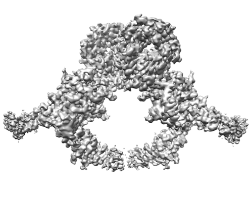 emd_21442.png emd_21442.png | 67.5 KB | ||
| Filedesc metadata | emd-21442.cif.gz | 7.2 KB | ||
| Others | emd_21442_additional.map.gz | 8.1 MB | ||
| Archive directory |  http://ftp.pdbj.org/pub/emdb/structures/EMD-21442ftp://ftp.pdbj.org/pub/emdb/structures/EMD-21442 http://ftp.pdbj.org/pub/emdb/structures/EMD-21442ftp://ftp.pdbj.org/pub/emdb/structures/EMD-21442 | HTTPS FTP |
-Related structure data
| Related structure data |  6vxkMC M: atomic model generated by this map C: citing same article ( |
|---|---|
| Similar structure data |
-Links
| EMDB pages | EMDB (EBI/PDBe) / EMDataResource |
|---|---|
| Related items in Molecule of the Month |


-Map
| File | Download / File: emd_21442.map.gz / Format: CCP4 / Size: 178 MB / Type: IMAGE STORED AS FLOATING POINT NUMBER (4 BYTES) | ||||||||||||||||||||||||||||||||||||||||||||||||||||||||||||||||||||
|---|---|---|---|---|---|---|---|---|---|---|---|---|---|---|---|---|---|---|---|---|---|---|---|---|---|---|---|---|---|---|---|---|---|---|---|---|---|---|---|---|---|---|---|---|---|---|---|---|---|---|---|---|---|---|---|---|---|---|---|---|---|---|---|---|---|---|---|---|---|
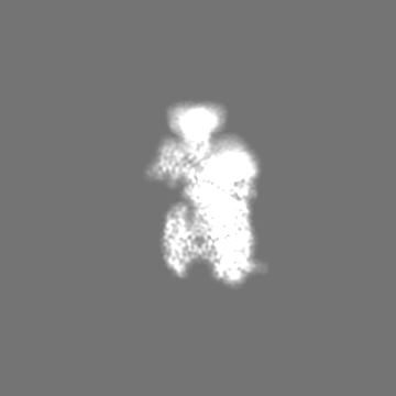
| Annotation | Dimeric map | ||||||||||||||||||||||||||||||||||||||||||||||||||||||||||||||||||||
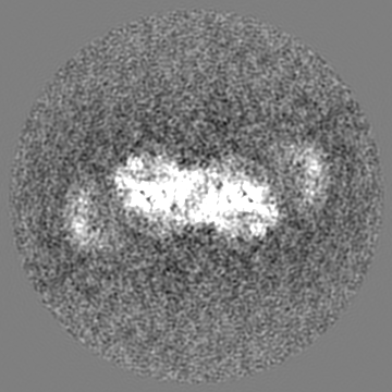
| Projections & slices | Image control
Images are generated by Spider. | ||||||||||||||||||||||||||||||||||||||||||||||||||||||||||||||||||||
| Voxel size | X=Y=Z: 0.828 Å | ||||||||||||||||||||||||||||||||||||||||||||||||||||||||||||||||||||
| Density |
| ||||||||||||||||||||||||||||||||||||||||||||||||||||||||||||||||||||
| Symmetry | Space group: 1 | ||||||||||||||||||||||||||||||||||||||||||||||||||||||||||||||||||||
| Details | EMDB XML:
CCP4 map header:
| ||||||||||||||||||||||||||||||||||||||||||||||||||||||||||||||||||||
 Z (Sec.)
 Y (Row.)
 X (Col.)


-Supplemental data
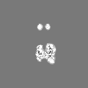
-Additional map: Focused refined half map
| File | emd_21442_additional.map | ||||||||||||
|---|---|---|---|---|---|---|---|---|---|---|---|---|---|
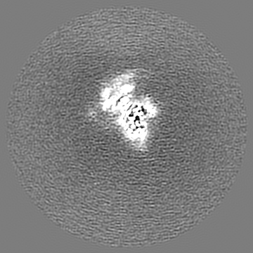
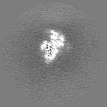
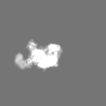
| Annotation | Focused refined half map | ||||||||||||
| Projections & Slices |
| ||||||||||||
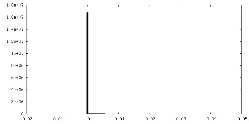
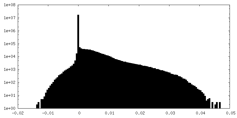
| Density Histograms |

- Sample components
-Entire : Complex between A39R and PlexinC1
| Entire | Name: Complex between A39R and PlexinC1 |
|---|---|
| Components |
|
-Supramolecule #1: Complex between A39R and PlexinC1
| Supramolecule | Name: Complex between A39R and PlexinC1 / type: complex / ID: 1 / Parent: 0 / Macromolecule list: #1-#2 / Details: Full-length PlexinC1 in complex with A39R |
|---|---|
| Source (natural) | Organism: Homo sapiens (human) |
| Molecular weight | Theoretical: 400 KDa |
-Supramolecule #2: A39R dimer
| Supramolecule | Name: A39R dimer / type: complex / ID: 2 / Parent: 1 / Macromolecule list: #1 |
|---|---|
| Source (natural) | Organism: Ectromelia virus |
-Supramolecule #3: PlexinC1
| Supramolecule | Name: PlexinC1 / type: complex / ID: 3 / Parent: 1 / Macromolecule list: #2 |
|---|---|
| Source (natural) | Organism: Homo sapiens (human) |
-Macromolecule #1: Semaphorin-like protein 139
| Macromolecule | Name: Semaphorin-like protein 139 / type: protein_or_peptide / ID: 1 / Number of copies: 2 / Enantiomer: LEVO |
|---|---|
| Source (natural) | Organism: Ectromelia virus (strain Moscow) / Strain: Moscow |
| Molecular weight | Theoretical: 45.330195 KDa |
| Recombinant expression | Organism: Homo sapiens (human) |
| Sequence | String: ELEIEWHKFE TSEEIISTYL IDDVLYTGVN GAVYTFSNNE LNKTGLTNNN NYITTSIKVE DTLVCGTNNG NPKCWKIDGS EDPKYRGRG YAPYQNSKVT IISHNECVLS DINISKEGIK RWRRFDGPCG YDLYTADNVI PKDGVRGAFV DKDGTYDKVY I LFTDTIDT ...String: ELEIEWHKFE TSEEIISTYL IDDVLYTGVN GAVYTFSNNE LNKTGLTNNN NYITTSIKVE DTLVCGTNNG NPKCWKIDGS EDPKYRGRG YAPYQNSKVT IISHNECVLS DINISKEGIK RWRRFDGPCG YDLYTADNVI PKDGVRGAFV DKDGTYDKVY I LFTDTIDT KRIVKIPYIA QMCLNDEGGP SSLSSHRWST FLKVELECDI DGRSYRQIIH SKAIKTDNDT ILYVFFDSPY SK SALCTYS MNAIKHSFST SKLGGYTKQL PSPAPGICLP AGKVVPHTTF DIIEQYNELD DIIKPLSQPI FEGPSGVKWF DIK EKENEH REYRIYFIKE NTIYSFDTKS KQTRSAQVDA RLFSVMVTSK PLFIADIGIG VGIPRMKKIL KMGTHHHHHH HH UniProtKB: Semaphorin-like protein 139 |
-Macromolecule #2: Plexin-C1
| Macromolecule | Name: Plexin-C1 / type: protein_or_peptide / ID: 2 / Number of copies: 2 / Enantiomer: LEVO |
|---|---|
| Source (natural) | Organism: Homo sapiens (human) |
| Molecular weight | Theoretical: 173.487 KDa |
| Recombinant expression | Organism: Homo sapiens (human) |
| Sequence | String: ADEPVWRSEQ AIGAIAASQE DGVFVASGSC LDQLDYSLEH SLSRLYRDQA GNCTEPVSLA PPARPRPGSS FSKLLLPYRE GAAGLGGLL LTGWTFDRGA CEVRPLGNLS RNSLRNGTEV VSCHPQGSTA GVVYRAGRNN RWYLAVAATY VLPEPETASR C NPAASDHD ...String: ADEPVWRSEQ AIGAIAASQE DGVFVASGSC LDQLDYSLEH SLSRLYRDQA GNCTEPVSLA PPARPRPGSS FSKLLLPYRE GAAGLGGLL LTGWTFDRGA CEVRPLGNLS RNSLRNGTEV VSCHPQGSTA GVVYRAGRNN RWYLAVAATY VLPEPETASR C NPAASDHD TAIALKDTEG RSLATQELGR LKLCEGAGSL HFVDAFLWNG SIYFPYYPYN YTSGAATGWP SMARIAQSTE VL FQGQASL DCGHGHPDGR RLLLSSSLVE ALDVWAGVFS AAAGEGQERR SPTTTALCLF RMSEIQARAK RVSWDFKTAE SHC KEGDQP ERVQPIASST LIHSDLTSVY GTVVMNRTVL FLGTGDGQLL KVILGENLTS NCPEVIYEIK EETPVFYKLV PDPV KNIYI YLTAGKEVRR IRVANCNKHK SCSECLTATD PHCGWCHSLQ RCTFQGDCVH SENLENWLDI SSGAKKCPKI QIIRS SKEK TTVTMVGSFS PRHSKCMVKN VDSSRELCQN KSQPNRTCTC SIPTRATYKD VSVVNVMFSF GSWNLSDRFN FTNCSS LKE CPACVETGCA WCKSARRCIH PFTACDPSDY ERNQEQCPVA VEKTSGGGRP KENKGNRTNQ ALQVFYIKSI EPQKVST LG KSNVIVTGAN FTRASNITMI LKGTSTCDKD VIQVSHVLND THMKFSLPSS RKEMKDVCIQ FDGGNCSSVG SLSYIALP H CSLIFPATTW ISGGQNITMM GRNFDVIDNL IISHELKGNI NVSEYCVATY CGFLAPSLKS SKVRTNVTVK LRVQDTYLD CGTLQYREDP RFTGYRVESE VDTELEVKIQ KENDNFNISK KDIEITLFHG ENGQLNCSFE NITRNQDLTT ILCKIKGIKT ASTIANSSK KVRVKLGNLE LYVEQESVPS TWYFLIVLPV LLVIVIFAAV GVTRHKSKEL SRKQSQQLEL LESELRKEIR D GFAELQMD KLDVVDSFGT VPFLDYKHFA LRTFFPESGG FTHIFTEDMH NRDANDKNES LTALDALICN KSFLVTVIHT LE KQKNFSV KDRCLFASFL TIALQTKLVY LTSILEVLTR DLMEQCSNMQ PKLMLRRTES VVEKLLTNWM SVCLSGFLRE TVG EPFYLL VTTLNQKINK GPVDVITCKA LYTLNEDWLL WQVPEFSTVA LNVVFEKIPE NESADVCRNI SVNVLDCDTI GQAK EKIFQ AFLSKNGSPY GLQLNEIGLE LQMGTRQKEL LDIDSSSVIL EDGITKLNTI GHYEISNGST IKVFKKIANF TSDVE YSDD HCHLILPDSE AFQDVQGKRH RGKHKFKVKE MYLTKLLSTK VAIHSVLEKL FRSIWSLPNS RAPFAIKYFF DFLDAQ AEN KKITDPDVVH IWKTNSLPLR FWVNILKNPQ FVFDIKKTPH IDGCLSVIAQ AFMDAFSLTE QQLGKEAPTN KLLYAKD IP TYKEEVKSYY KAIRDLPPLS SSEMEEFLTQ ESKKHENEFN EEVALTEIYK YIVKYFDEIL NKLERERGLE EAQKQLLH V KVLFDEKKKC KWMGTSSGLE VLFQ UniProtKB: Plexin-C1 |
-Macromolecule #3: 2-acetamido-2-deoxy-beta-D-glucopyranose
| Macromolecule | Name: 2-acetamido-2-deoxy-beta-D-glucopyranose / type: ligand / ID: 3 / Number of copies: 18 / Formula: NAG |
|---|---|
| Molecular weight | Theoretical: 221.208 Da |
| Chemical component information |  ChemComp-NAG: |
-Experimental details
-Structure determination
| Method | cryo EM |
|---|---|
 Processing Processing | single particle reconstruction |
| Aggregation state | particle |
-Sample preparation
| Concentration | 0.5 mg/mL | |||||||||
|---|---|---|---|---|---|---|---|---|---|---|
| Buffer | pH: 7.5 Component:
| |||||||||
| Grid | Model: Quantifoil, UltrAuFoil, R1.2/1.3 / Support film - Material: CARBON / Support film - topology: HOLEY ARRAY / Pretreatment - Type: GLOW DISCHARGE | |||||||||
| Vitrification | Cryogen name: NITROGEN |
- Electron microscopy
| Microscope | FEI TITAN KRIOS |
|---|---|
| Image recording | Film or detector model: GATAN K3 BIOQUANTUM (6k x 4k) / Average electron dose: 2.0 e/Å2 |
| Electron beam | Acceleration voltage: 300 kV / Electron source:  FIELD EMISSION GUN FIELD EMISSION GUN |
| Electron optics | Illumination mode: FLOOD BEAM / Imaging mode: BRIGHT FIELD |
| Experimental equipment |  Model: Titan Krios / Image courtesy: FEI Company |
-Image processing
| Startup model | Type of model: INSILICO MODEL |
|---|---|
| Final reconstruction | Applied symmetry - Point group: C2 (2 fold cyclic) / Resolution.type: BY AUTHOR / Resolution: 3.1 Å / Resolution method: FSC 0.143 CUT-OFF / Number images used: 143750 |
| Initial angle assignment | Type: MAXIMUM LIKELIHOOD |
| Final angle assignment | Type: MAXIMUM LIKELIHOOD |