 Movie
Movie Controller

[English] 日本語
 Yorodumi
- PDB-6qx7: The cryo-EM structure of connector in bacteriophage phi29 prohead -
+ Open data

- Basic information
| Entry | Database: PDB / ID: 6qx7 | ||||||||||||
|---|---|---|---|---|---|---|---|---|---|---|---|---|---|
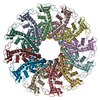
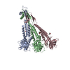
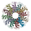
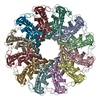
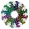
| Title | The cryo-EM structure of connector in bacteriophage phi29 prohead | ||||||||||||
 Components Components | Portal protein | ||||||||||||
 Keywords Keywords | VIRUS / bacteriophage / phi29 / prohead | ||||||||||||
| Function / homology | Portal protein Gp10 / Portal protein Gp10 superfamily / Phage Connector (GP10) / viral procapsid / viral portal complex / symbiont genome ejection through host cell envelope, short tail mechanism / viral DNA genome packaging / RNA binding / Portal protein Function and homology information Function and homology information | ||||||||||||
| Biological species |   Bacillus phage phi29 (virus) Bacillus phage phi29 (virus) | ||||||||||||
| Method | ELECTRON MICROSCOPY / single particle reconstruction / cryo EM / Resolution: 3.8 Å | ||||||||||||
 Authors Authors | Xu, J. / Gui, M. / Xiang, Y. | ||||||||||||
| Funding support |  China, 3items China, 3items
| ||||||||||||
 Citation Citation | Journal: Nat Commun / Year: 2019 Title: Structural assembly of the tailed bacteriophage ϕ29. Authors: Jingwei Xu / Dianhong Wang / Miao Gui / Ye Xiang /   Abstract: The mature virion of the tailed bacteriophage ϕ29 is an ~33 MDa complex that contains more than 450 subunits of seven structural proteins assembling into a prolate head and a short non-contractile ...The mature virion of the tailed bacteriophage ϕ29 is an ~33 MDa complex that contains more than 450 subunits of seven structural proteins assembling into a prolate head and a short non-contractile tail. Here, we report the near-atomic structures of the ϕ29 pre-genome packaging head (prohead), the mature virion and the genome-emptied virion. Structural comparisons suggest local rotation or oscillation of the head-tail connector upon DNA packaging and release. Termination of the DNA packaging occurs through pressure-dependent correlative positional and conformational changes in the connector. The funnel-shaped tail lower collar attaches the expanded narrow end of the connector and has a 180-Å long, 24-strand β barrel narrow stem tube that undergoes conformational changes upon genome release. The appendages form an interlocked assembly attaching the tail around the collar. The membrane active long loops at the distal end of the tail knob exit during the late stage of infection and form the cone-shaped tip of a largely hydrophobic helix barrel, prepared for membrane penetration. | ||||||||||||
| History |
|
- Structure visualization
| Movie |
Movie viewer |
|---|---|
| Structure viewer | Molecule: MolmilJmol/JSmol |
- Downloads & links
-Download
| PDBx/mmCIF format | 6qx7.cif.gz | 541 KB | Display | PDBx/mmCIF format |
|---|---|---|---|---|
| PDB format | pdb6qx7.ent.gz | Display | PDB format | |
| PDBx/mmJSON format | 6qx7.json.gz | Tree view | PDBx/mmJSON format | |
| Others |  Other downloads Other downloads |
-Validation report
| Summary document | 6qx7_validation.pdf.gz | 752.8 KB | Display | wwPDB validaton report |
|---|---|---|---|---|
| Full document | 6qx7_full_validation.pdf.gz | 763.7 KB | Display | |
| Data in XML | 6qx7_validation.xml.gz | 75.2 KB | Display | |
| Data in CIF | 6qx7_validation.cif.gz | 100.8 KB | Display | |
| Arichive directory | https://data.pdbj.org/pub/pdb/validation_reports/qx/6qx7ftp://data.pdbj.org/pub/pdb/validation_reports/qx/6qx7 | HTTPS FTP |
-Related structure data
| Related structure data | 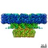 4662MC 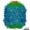 4655C 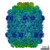 4677C 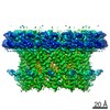 4678C 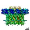 4679C 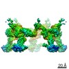 4680C 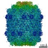 4681C 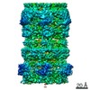 4682C 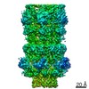 4683C 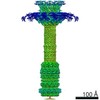 4684C 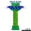 4685C 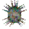 6qvkC  6qydC  6qyjC  6qymC  6qyyC 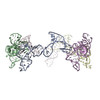 6qyzC 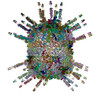 6qz0C 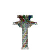 6qz9C 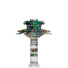 6qzfC M: map data used to model this data C: citing same article ( |
|---|---|
| Similar structure data |
-Links
 PDBj
PDBj- Assembly
| Deposited unit | 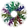
|
|---|---|
| 1 |
|
-Components
| #1: Protein | Mass: 35917.293 Da / Num. of mol.: 12 Source method: isolated from a genetically manipulated source Source: (gene. exp.) Bacillus phage phi29 (virus) / Gene: 10 / Production host: Bacillus phage phi29 (virus) / References: UniProt: P04332 |
|---|
-Experimental details
-Experiment
| Experiment | Method: ELECTRON MICROSCOPY |
|---|---|
| EM experiment | Aggregation state: PARTICLE / 3D reconstruction method: single particle reconstruction |
- Sample preparation
| Component | Name: Bacillus phage phi29 / Type: VIRUS / Entity ID: all / Source: NATURAL |
|---|---|
| Source (natural) | Organism: Bacillus phage phi29 (virus) |
| Details of virus | Empty: YES / Enveloped: NO / Isolate: STRAIN / Type: PRION |
| Buffer solution | pH: 7.5 |
| Specimen | Embedding applied: NO / Shadowing applied: NO / Staining applied: NO / Vitrification applied: YES |
| Vitrification | Cryogen name: ETHANE |
- Electron microscopy imaging
| Experimental equipment |  Model: Titan Krios / Image courtesy: FEI Company |
|---|---|
| Microscopy | Model: FEI TITAN KRIOS |
| Electron gun | Electron source:  FIELD EMISSION GUN / Accelerating voltage: 300 kV / Illumination mode: FLOOD BEAM FIELD EMISSION GUN / Accelerating voltage: 300 kV / Illumination mode: FLOOD BEAM |
| Electron lens | Mode: BRIGHT FIELD |
| Image recording | Electron dose: 25 e/Å2 / Film or detector model: KODAK SO-163 FILM |
- Processing
| CTF correction | Type: PHASE FLIPPING ONLY |
|---|---|
| 3D reconstruction | Resolution: 3.8 Å / Resolution method: FSC 0.143 CUT-OFF / Num. of particles: 18230 / Symmetry type: POINT |
