National Institutes of Health/National Institute Of Allergy and Infectious Diseases (NIH/NIAID)
AI-100645
米国
National Institutes of Health/National Institute Of Allergy and Infectious Diseases (NIH/NIAID)
AI129721
米国
引用
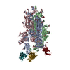
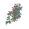
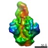
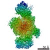
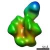
ジャーナル: J Mol Biol / 年: 2020 タイトル: Cryo-EM Structure of Full-length HIV-1 Env Bound With the Fab of Antibody PG16. 著者: Junhua Pan / Hanqin Peng / Bing Chen / Stephen C Harrison / 要旨: The HIV-1 envelope protein (Env) is the target of neutralizing antibodies and the template for vaccine immunogen design. The dynamic conformational equilibrium of trimeric Env influences its ...The HIV-1 envelope protein (Env) is the target of neutralizing antibodies and the template for vaccine immunogen design. The dynamic conformational equilibrium of trimeric Env influences its antigenicity and potential immunogenicity. Antibodies that bind at the trimer apex stabilize a "closed" conformation characteristic of the most difficult to neutralize isolates. A goal of vaccine development is therefore to mimic the closed conformation in a designed immunogen. A disulfide-stabilized, trimeric Env ectodomain-the "SOSIP" construct-has many of the relevant properties; it is also particularly suitable for structure determination. Some single-molecule studies have, however, suggested that the SOSIP trimer is not a good representation of Env on the surface of a virion or an infected cell. We isolated Env (fully cleaved to gp120 and gp41) from the surface of expressing cells using tagged, apex-binding Fab PG16 and determined the structure of the PG16-Env complex by cryo-EM to an overall resolution of 4.6 Å. Placing the only purification tag on the Fab ensured that the isolated Env was continuously stabilized in its closed, native conformation. The Env structure in this complex corresponds closely to the SOSIP structures determined by both x-ray crystallography and cryo-EM. Although the membrane-interacting elements are not resolved in our reconstruction, we can make inferences about the connection between ectodomain and membrane-proximal external region (MPER) by reference to the published cryo-tomography structure of an Env "spike" and the NMR structure of the MPER-transmembrane segment. We discuss these results in view of the conflicting interpretations in the literature.
モード: BRIGHT FIELD / 倍率(公称値): 29000 X / 倍率(補正後): 30488 X / 最大 デフォーカス(公称値): 3000 nm / 最小 デフォーカス(公称値): 1000 nm / Calibrated defocus min: 800 nm / Cs: 2.26 mm / C2レンズ絞り径: 50 µm / アライメント法: COMA FREE
試料ホルダ
凍結剤: NITROGEN / 試料ホルダーモデル: OTHER / 最高温度: 70 K / 最低温度: 70 K
撮影
平均露光時間: 0.125 sec. / 電子線照射量: 40 e/Å2 / 検出モード: SUPER-RESOLUTION フィルム・検出器のモデル: GATAN K2 SUMMIT (4k x 4k) 撮影したグリッド数: 2 / 実像数: 8138 詳細: image size and sampling interval reported here are for super resolution. The physical pixel size of the camera is 5.0 um and image size is 3838 pixels by 3710 pixels.
 ムービー
ムービー コントローラー


 データを開く
 基本情報
基本情報 要素
要素 キーワード
キーワード 機能・相同性情報
 Human immunodeficiency virus 1 (ヒト免疫不全ウイルス)
 Homo sapiens (ヒト)
 データ登録者
 米国, 2件
  引用
引用 構造の表示
構造の表示 ダウンロードとリンク
 その他のダウンロード


 PDBj
PDBj


 集合体
集合体

 タイプ: D-saccharide, beta linking / 分子量: 221.208 Da / 分子数: 44 / 由来タイプ: 組換発現 / 式: C8H15NO6
 試料調製
試料調製 電子顕微鏡撮影
 FIELD EMISSION GUN / 加速電圧: 300 kV / 照射モード: SPOT SCAN
 解析
解析