National Institutes of Health/National Human Genome Research Institute (NIH/NHGRI)
R35 GM122588
United States
Citation
Journal: Structure / Year: 2019 Title: Collection of Continuous Rotation MicroED Data from Ion Beam-Milled Crystals of Any Size. Authors: Michael W Martynowycz / Wei Zhao / Johan Hattne / Grant J Jensen / Tamir Gonen / Abstract: Microcrystal electron diffraction (MicroED) allows for macromolecular structure solution from nanocrystals. To create crystals of suitable size for MicroED data collection, sample preparation ...Microcrystal electron diffraction (MicroED) allows for macromolecular structure solution from nanocrystals. To create crystals of suitable size for MicroED data collection, sample preparation typically involves sonication or pipetting a slurry of crystals from a crystallization drop. The resultant crystal fragments are fragile and the quality of the data that can be obtained from them is sensitive to subsequent sample preparation for cryoelectron microscopy as interactions in the water-air interface can damage crystals during blotting. Here, we demonstrate the use of a focused ion beam to generate lamellae of macromolecular protein crystals for continuous rotation MicroED that are of ideal thickness, easy to locate, and require no blotting optimization. In this manner, crystals of nearly any size may be scooped and milled to desired dimensions prior to data collection, thus streamlining the methodology for sample preparation for MicroED.
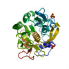
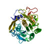
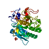
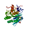
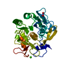
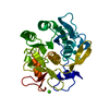
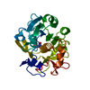
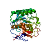
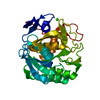
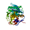
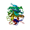
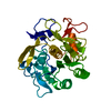
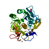
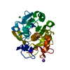
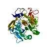
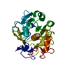
ProteinaseK / Endopeptidase K / Tritirachium alkaline proteinase
Mass: 28958.791 Da / Num. of mol.: 1 Source method: isolated from a genetically manipulated source Source: (gene. exp.) Engyodontium album (fungus) / Gene: PROK / Production host: Engyodontium album (fungus) / References: UniProt: P06873, peptidase K
Cryogen: NITROGEN / Specimen holder model: FEI TITAN KRIOS AUTOGRID HOLDER / Temperature (max): 90 K / Temperature (min): 77 K
Image recording
Average exposure time: 2 sec. / Electron dose: 0.04 e/Å2 / Film or detector model: FEI CETA (4k x 4k) / Num. of diffraction images: 100 / Num. of grids imaged: 1 Details: Continuous rotation from -30 to +30 with a rotation rate of 0.2 degrees per second and a readout every 3 seconds.
Image scans
Sampling size: 28 µm / Width: 4096 / Height: 4096
EM diffraction
Camera length: 1700 mm
EM diffraction shell
Resolution: 2.75→3.1483 Å / Fourier space coverage: 88.23 % / Multiplicity: 2.9 / Num. of structure factors: 1831 / Phase residual: 28.11 °
EM diffraction stats
Fourier space coverage: 88.4 % / High resolution: 2.75 Å / Num. of intensities measured: 31013 / Num. of structure factors: 10807 / Phase error: 22.41 ° / Phase residual: 22.41 ° / Phase error rejection criteria: 0 / Rmerge: 0.47 / Rsym: 0.39
In the structure databanks used in Yorodumi, some data are registered as the other names, "COVID-19 virus" and "2019-nCoV". Here are the details of the virus and the list of structure data.
Jan 31, 2019. EMDB accession codes are about to change! (news from PDBe EMDB page)
EMDB accession codes are about to change! (news from PDBe EMDB page)
The allocation of 4 digits for EMDB accession codes will soon come to an end. Whilst these codes will remain in use, new EMDB accession codes will include an additional digit and will expand incrementally as the available range of codes is exhausted. The current 4-digit format prefixed with “EMD-” (i.e. EMD-XXXX) will advance to a 5-digit format (i.e. EMD-XXXXX), and so on. It is currently estimated that the 4-digit codes will be depleted around Spring 2019, at which point the 5-digit format will come into force.
The EM Navigator/Yorodumi systems omit the EMD- prefix.
Related info.:Q: What is EMD? / ID/Accession-code notation in Yorodumi/EM Navigator
Yorodumi is a browser for structure data from EMDB, PDB, SASBDB, etc.
This page is also the successor to EM Navigator detail page, and also detail information page/front-end page for Omokage search.
The word "yorodu" (or yorozu) is an old Japanese word meaning "ten thousand". "mi" (miru) is to see.
Related info.:EMDB / PDB / SASBDB / Comparison of 3 databanks / Yorodumi Search / Aug 31, 2016. New EM Navigator & Yorodumi / Yorodumi Papers / Jmol/JSmol / Function and homology information / Changes in new EM Navigator and Yorodumi
 Movie
Movie Controller

 Yorodumi
 Open data
 Basic information
 Components
 Keywords
 Function and homology information
 Engyodontium album (fungus)
 MOLECULAR REPLACEMENT / cryo EM / Resolution: 2.75 Å
  Authors
 United States, 1items
  Citation
 Structure visualization
 Downloads & links
 Other downloads


 PDBj
PDBj

 Assembly
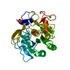

 Mass: 40.078 Da / Num. of mol.: 2 / Source method: obtained synthetically / Formula: Ca
 Mass: 96.063 Da / Num. of mol.: 1 / Source method: obtained synthetically / Formula: SO4
 Mass: 18.015 Da / Num. of mol.: 23 / Source method: isolated from a natural source / Formula: H2O
 Sample preparation
 Processing