[English] 日本語
 Yorodumi
- PDB-6n0f: Cryo-EM structure of the HO BMC shell: subregion classified for B... -
+ Open data
- Basic information
| Entry | Database: PDB / ID: 6n0f | |||||||||
|---|---|---|---|---|---|---|---|---|---|---|
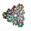
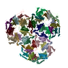
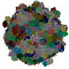
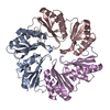
| Title | Cryo-EM structure of the HO BMC shell: subregion classified for BMC-T: TD-TSTSTS | |||||||||
 Components Components | (Microcompartments protein) x 3 | |||||||||
 Keywords Keywords | STRUCTURAL PROTEIN / microcompartment / shell / compartmentalization / BMC fold | |||||||||
| Function / homology |  Function and homology information Function and homology information | |||||||||
| Biological species |  Haliangium ochraceum (bacteria) Haliangium ochraceum (bacteria) | |||||||||
| Method | ELECTRON MICROSCOPY / single particle reconstruction / cryo EM / Resolution: 3.9 Å | |||||||||
 Authors Authors | Greber, B.J. / Sutter, M. / Kerfeld, C.A. | |||||||||
| Funding support |  United States, 2items United States, 2items
| |||||||||
 Citation Citation |  Journal: Structure / Year: 2019 Journal: Structure / Year: 2019Title: The Plasticity of Molecular Interactions Governs Bacterial Microcompartment Shell Assembly. Authors: Basil J Greber / Markus Sutter / Cheryl A Kerfeld /  Abstract: Bacterial microcompartments (BMCs) are composed of an enzymatic core encapsulated by a selectively permeable protein shell that enhances catalytic efficiency. Many pathogenic bacteria derive ...Bacterial microcompartments (BMCs) are composed of an enzymatic core encapsulated by a selectively permeable protein shell that enhances catalytic efficiency. Many pathogenic bacteria derive competitive advantages from their BMC-based catabolism, implicating BMCs as drug targets. BMC shells are of interest for bioengineering due to their diverse and selective permeability properties and because they self-assemble. A complete understanding of shell composition and organization is a prerequisite for biotechnological applications. Here, we report the cryoelectron microscopy structure of a BMC shell at 3.0-Å resolution, using an image-processing strategy that allowed us to determine the previously uncharacterized structural details of the interactions formed by the BMC-T and BMC-T shell subunits in the context of the assembled shell. We found unexpected structural plasticity among these interactions, resulting in distinct shell populations assembled from varying numbers of the BMC-T and BMC-T subunits. We discuss the implications of these findings on shell assembly and function. | |||||||||
| History |
|
- Structure visualization
| Movie |
 Movie viewer Movie viewer |
|---|---|
| Structure viewer | Molecule:  Molmil Molmil Jmol/JSmol Jmol/JSmol |
- Downloads & links
- Download
| PDBx/mmCIF format |  6n0f.cif.gz 6n0f.cif.gz | 968.9 KB | Display |  PDBx/mmCIF format PDBx/mmCIF format |
|---|---|---|---|---|
| PDB format |  pdb6n0f.ent.gz pdb6n0f.ent.gz | Display |  PDB format PDB format | |
| PDBx/mmJSON format |  6n0f.json.gz 6n0f.json.gz | Tree view |  PDBx/mmJSON format PDBx/mmJSON format | |
| Others |  Other downloads Other downloads |
-Validation report
| Summary document |  6n0f_validation.pdf.gz 6n0f_validation.pdf.gz | 1019.6 KB | Display |  wwPDB validaton report wwPDB validaton report |
|---|---|---|---|---|
| Full document |  6n0f_full_validation.pdf.gz 6n0f_full_validation.pdf.gz | 1.1 MB | Display | |
| Data in XML |  6n0f_validation.xml.gz 6n0f_validation.xml.gz | 147.7 KB | Display | |
| Data in CIF |  6n0f_validation.cif.gz 6n0f_validation.cif.gz | 233 KB | Display | |
| Arichive directory |  https://data.pdbj.org/pub/pdb/validation_reports/n0/6n0f https://data.pdbj.org/pub/pdb/validation_reports/n0/6n0f ftp://data.pdbj.org/pub/pdb/validation_reports/n0/6n0f ftp://data.pdbj.org/pub/pdb/validation_reports/n0/6n0f | HTTPS FTP |
-Related structure data
| Related structure data |  9314MC  9296C  9307C  9308C  9309C  9310C  9311C  9312C  9313C  9315C  6mzuC  6mzvC  6mzxC  6mzyC  6n06C  6n07C  6n09C  6n0gC M: map data used to model this data C: citing same article ( |
|---|---|
| Similar structure data |
- Links
Links
- Assembly
| Deposited unit | 
|
|---|---|
| 1 |
|
- Components
| #1: Protein | Mass: 22904.137 Da / Num. of mol.: 6 Source method: isolated from a genetically manipulated source Source: (gene. exp.)  Haliangium ochraceum (strain DSM 14365 / JCM 11303 / SMP-2) (bacteria) Haliangium ochraceum (strain DSM 14365 / JCM 11303 / SMP-2) (bacteria)Strain: DSM 14365 / JCM 11303 / SMP-2 / Gene: Hoch_5816 / Production host:  #2: Protein | Mass: 10126.718 Da / Num. of mol.: 36 Source method: isolated from a genetically manipulated source Source: (gene. exp.)  Haliangium ochraceum (strain DSM 14365 / JCM 11303 / SMP-2) (bacteria) Haliangium ochraceum (strain DSM 14365 / JCM 11303 / SMP-2) (bacteria)Strain: DSM 14365 / JCM 11303 / SMP-2 / Gene: Hoch_5815 / Production host:  #3: Protein | Mass: 21923.199 Da / Num. of mol.: 9 Source method: isolated from a genetically manipulated source Source: (gene. exp.)  Haliangium ochraceum (strain DSM 14365 / JCM 11303 / SMP-2) (bacteria) Haliangium ochraceum (strain DSM 14365 / JCM 11303 / SMP-2) (bacteria)Strain: DSM 14365 / JCM 11303 / SMP-2 / Gene: Hoch_5812 / Production host:  |
|---|
-Experimental details
-Experiment
| Experiment | Method: ELECTRON MICROSCOPY |
|---|---|
| EM experiment | Aggregation state: PARTICLE / 3D reconstruction method: single particle reconstruction |
- Sample preparation
| Component | Name: Bacterial microcompartment shell from Haliangium ochraceum Type: ORGANELLE OR CELLULAR COMPONENT / Entity ID: all / Source: RECOMBINANT | ||||||||||||||||||||
|---|---|---|---|---|---|---|---|---|---|---|---|---|---|---|---|---|---|---|---|---|---|
| Molecular weight | Value: 6.5 MDa / Experimental value: NO | ||||||||||||||||||||
| Source (natural) | Organism:  Haliangium ochraceum (strain DSM 14365 / JCM 11303 / SMP-2) (bacteria) Haliangium ochraceum (strain DSM 14365 / JCM 11303 / SMP-2) (bacteria)Strain: DSM 14365 / JCM 11303 / SMP-2 | ||||||||||||||||||||
| Source (recombinant) | Organism:  | ||||||||||||||||||||
| Buffer solution | pH: 7.4 | ||||||||||||||||||||
| Buffer component |
| ||||||||||||||||||||
| Specimen | Conc.: 3 mg/ml / Embedding applied: NO / Shadowing applied: NO / Staining applied: NO / Vitrification applied: YES | ||||||||||||||||||||
| Specimen support | Details: unspecified | ||||||||||||||||||||
| Vitrification | Instrument: FEI VITROBOT MARK IV / Cryogen name: ETHANE / Humidity: 100 % / Chamber temperature: 277 K Details: 5-7 seconds incubation of the sample on the grid before blotting and plunging |
- Electron microscopy imaging
| Microscopy | Model: FEI TITAN |
|---|---|
| Electron gun | Electron source:  FIELD EMISSION GUN / Accelerating voltage: 300 kV / Illumination mode: FLOOD BEAM FIELD EMISSION GUN / Accelerating voltage: 300 kV / Illumination mode: FLOOD BEAM |
| Electron lens | Mode: BRIGHT FIELD / Calibrated magnification: 48543 X / Calibrated defocus min: 1000 nm / Calibrated defocus max: 3500 nm / Cs: 2.7 mm / C2 aperture diameter: 50 µm / Alignment procedure: COMA FREE |
| Specimen holder | Cryogen: NITROGEN Specimen holder model: GATAN 626 SINGLE TILT LIQUID NITROGEN CRYO TRANSFER HOLDER |
| Image recording | Average exposure time: 4.5 sec. / Electron dose: 25 e/Å2 / Detector mode: COUNTING / Film or detector model: GATAN K2 SUMMIT (4k x 4k) / Num. of grids imaged: 1 / Num. of real images: 928 Details: 928 images retained after inspection for image quality. |
| Image scans | Sampling size: 5 µm / Width: 3838 / Height: 3710 / Movie frames/image: 30 / Used frames/image: 1-30 |
- Processing
| EM software |
| ||||||||||||||||||||||||||||||||||||
|---|---|---|---|---|---|---|---|---|---|---|---|---|---|---|---|---|---|---|---|---|---|---|---|---|---|---|---|---|---|---|---|---|---|---|---|---|---|
| CTF correction | Details: Initial CTF fitting using CTFFIND4, CTF correction applied within RELION Type: PHASE FLIPPING AND AMPLITUDE CORRECTION | ||||||||||||||||||||||||||||||||||||
| Particle selection | Num. of particles selected: 31800 Details: 1000 particles were picked manually to generate reference templates for subsequent auto-picking in RELION 1.4. | ||||||||||||||||||||||||||||||||||||
| Symmetry | Point symmetry: C1 (asymmetric) | ||||||||||||||||||||||||||||||||||||
| 3D reconstruction | Resolution: 3.9 Å / Resolution method: FSC 0.143 CUT-OFF / Num. of particles: 25580 / Algorithm: FOURIER SPACE Details: The selected particle subset was refined without masking and subsequently masked to reveal only the subregion of the BMC shell to which the focused classification had been applied. Num. of class averages: 1 / Symmetry type: POINT | ||||||||||||||||||||||||||||||||||||
| Atomic model building | Protocol: OTHER / Space: REAL | ||||||||||||||||||||||||||||||||||||
| Atomic model building |
|
 Movie
Movie Controller


 PDBj
PDBj


