 ムービー
ムービー コントローラー


+ データを開く

- 基本情報
基本情報
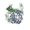
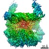
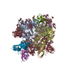
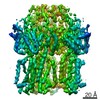
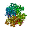
| 登録情報 | データベース: PDB / ID: 6klo | |||||||||
|---|---|---|---|---|---|---|---|---|---|---|
| タイトル | Complex structure of Iota toxin enzymatic component (Ia) and binding component (Ib) pore with short stem | |||||||||
 要素 要素 |
| |||||||||
 キーワード キーワード | TOXIN / Bacterial binary toxin / Protein translocation channel | |||||||||
| 機能・相同性 |  機能・相同性情報 機能・相同性情報 | |||||||||
| 生物種 |   Clostridium perfringens (ウェルシュ菌) Clostridium perfringens (ウェルシュ菌) | |||||||||
| 手法 | 電子顕微鏡法 / 単粒子再構成法 / クライオ電子顕微鏡法 / 解像度: 2.8 Å | |||||||||
 データ登録者 データ登録者 | Yoshida, T. / Yamada, T. / Kawamoto, A. / Mitsuoka, K. / Iwasaki, K. / Tsuge, H. | |||||||||
| 資金援助 |  日本, 2件 日本, 2件
| |||||||||
 引用 引用 | ジャーナル: Nat Struct Mol Biol / 年: 2020 タイトル: Cryo-EM structures reveal translocational unfolding in the clostridial binary iota toxin complex. 著者: Tomohito Yamada / Toru Yoshida / Akihiro Kawamoto / Kaoru Mitsuoka / Kenji Iwasaki / Hideaki Tsuge / 要旨: The iota toxin produced by Clostridium perfringens type E is a binary toxin comprising two independent polypeptides: Ia, an ADP-ribosyltransferase, and Ib, which is involved in cell binding and ...The iota toxin produced by Clostridium perfringens type E is a binary toxin comprising two independent polypeptides: Ia, an ADP-ribosyltransferase, and Ib, which is involved in cell binding and translocation of Ia across the cell membrane. Here we report cryo-EM structures of the translocation channel Ib-pore and its complex with Ia. The high-resolution Ib-pore structure demonstrates a similar structural framework to that of the catalytic ϕ-clamp of the anthrax protective antigen pore. However, the Ia-bound Ib-pore structure shows a unique binding mode of Ia: one Ia binds to the Ib-pore, and the Ia amino-terminal domain forms multiple weak interactions with two additional Ib-pore constriction sites. Furthermore, Ib-binding induces tilting and partial unfolding of the Ia N-terminal α-helix, permitting its extension to the ϕ-clamp gate. This new mechanism of N-terminal unfolding is crucial for protein translocation. | |||||||||
| 履歴 |
|
- 構造の表示
構造の表示
| ムービー |
ムービービューア |
|---|---|
| 構造ビューア | 分子: MolmilJmol/JSmol |
- ダウンロードとリンク
-ダウンロード
| PDBx/mmCIF形式 | 6klo.cif.gz | 535.9 KB | 表示 | PDBx/mmCIF形式 |
|---|---|---|---|---|
| PDB形式 | pdb6klo.ent.gz | 427.3 KB | 表示 | PDB形式 |
| PDBx/mmJSON形式 | 6klo.json.gz | ツリー表示 | PDBx/mmJSON形式 | |
| その他 |  その他のダウンロード その他のダウンロード |
-検証レポート
| 文書・要旨 | 6klo_validation.pdf.gz | 795 KB | 表示 | wwPDB検証レポート |
|---|---|---|---|---|
| 文書・詳細版 | 6klo_full_validation.pdf.gz | 814.2 KB | 表示 | |
| XML形式データ | 6klo_validation.xml.gz | 80.2 KB | 表示 | |
| CIF形式データ | 6klo_validation.cif.gz | 123.6 KB | 表示 | |
| アーカイブディレクトリ | https://data.pdbj.org/pub/pdb/validation_reports/kl/6kloftp://data.pdbj.org/pub/pdb/validation_reports/kl/6klo | HTTPS FTP |
-関連構造データ


-リンク
 PDBj
PDBj
- 集合体
集合体
| 登録構造単位 | 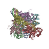
|
|---|---|
| 1 |
|
-要素
| #1: タンパク質 | 分子量: 74378.820 Da / 分子数: 7 / 由来タイプ: 組換発現 由来: (組換発現) Clostridium perfringens (ウェルシュ菌)プラスミド: pGEX-4T-1 / 発現宿主: #2: タンパク質 | | 分子量: 48061.988 Da / 分子数: 1 / 由来タイプ: 組換発現 由来: (組換発現) Clostridium perfringens (ウェルシュ菌)プラスミド: pET15b / 発現宿主: #3: 化合物 | ChemComp-CA /   分子量: 40.078 Da / 分子数: 14 / 由来タイプ: 合成 / 式: Ca 分子量: 40.078 Da / 分子数: 14 / 由来タイプ: 合成 / 式: Ca研究の焦点であるリガンドがあるか | N | |
|---|
-実験情報
-実験
| 実験 | 手法: 電子顕微鏡法 |
|---|---|
| EM実験 | 試料の集合状態: PARTICLE / 3次元再構成法: 単粒子再構成法 |
- 試料調製
試料調製
| 構成要素 | 名称: Complex structure of Iota toxin enzymatic component (Ia) and binding component (Ib) pore with short stem タイプ: COMPLEX / Entity ID: #1-#2 / 由来: RECOMBINANT | ||||||||||||||||||||
|---|---|---|---|---|---|---|---|---|---|---|---|---|---|---|---|---|---|---|---|---|---|
| 分子量 | 値: 0.57 MDa / 実験値: NO | ||||||||||||||||||||
| 由来(天然) | 生物種: Clostridium perfringens (ウェルシュ菌) | ||||||||||||||||||||
| 由来(組換発現) | 生物種: | ||||||||||||||||||||
| 緩衝液 | pH: 7.5 | ||||||||||||||||||||
| 緩衝液成分 |
| ||||||||||||||||||||
| 試料 | 濃度: 0.48 mg/ml / 包埋: NO / シャドウイング: NO / 染色: NO / 凍結: YES | ||||||||||||||||||||
| 試料支持 | グリッドの材料: COPPER / グリッドのサイズ: 200 divisions/in. / グリッドのタイプ: Quantifoil R1.2/1.3 | ||||||||||||||||||||
| 急速凍結 | 装置: FEI VITROBOT MARK IV / 凍結剤: ETHANE / 湿度: 100 % / 凍結前の試料温度: 277 K |
- 電子顕微鏡撮影
| 実験機器 |  モデル: Titan Krios / 画像提供: FEI Company |
|---|---|
| 顕微鏡 | モデル: FEI TITAN KRIOS |
| 電子銃 | 電子線源:  FIELD EMISSION GUN / 加速電圧: 300 kV / 照射モード: FLOOD BEAM FIELD EMISSION GUN / 加速電圧: 300 kV / 照射モード: FLOOD BEAM |
| 電子レンズ | モード: BRIGHT FIELD / 倍率(公称値): 59000 X / 最大 デフォーカス(公称値): 2500 nm / 最小 デフォーカス(公称値): 800 nm / C2レンズ絞り径: 50 µm |
| 試料ホルダ | 凍結剤: NITROGEN 試料ホルダーモデル: FEI TITAN KRIOS AUTOGRID HOLDER 最高温度: 78.9 K / 最低温度: 78.9 K |
| 撮影 | 平均露光時間: 84.09 sec. / 電子線照射量: 50 e/Å2 / 検出モード: COUNTING フィルム・検出器のモデル: FEI FALCON III (4k x 4k) 撮影したグリッド数: 1 / 実像数: 2151 |
| 電子光学装置 | 球面収差補正装置: Microscope was modified with a Cs corrector |
| 画像スキャン | 横: 4096 / 縦: 4096 |
- 解析
解析
| ソフトウェア | 名称: PHENIX / バージョン: (1.15.2_3472: phenix.real_space_refine) / 分類: 精密化 | ||||||||||||||||||||||||||||||||||||||||
|---|---|---|---|---|---|---|---|---|---|---|---|---|---|---|---|---|---|---|---|---|---|---|---|---|---|---|---|---|---|---|---|---|---|---|---|---|---|---|---|---|---|
| EMソフトウェア |
| ||||||||||||||||||||||||||||||||||||||||
| CTF補正 | タイプ: PHASE FLIPPING AND AMPLITUDE CORRECTION | ||||||||||||||||||||||||||||||||||||||||
| 粒子像の選択 | 選択した粒子像数: 871264 | ||||||||||||||||||||||||||||||||||||||||
| 対称性 | 点対称性: C1 (非対称) | ||||||||||||||||||||||||||||||||||||||||
| 3次元再構成 | 解像度: 2.8 Å / 解像度の算出法: FSC 0.143 CUT-OFF / 粒子像の数: 135359 / 対称性のタイプ: POINT | ||||||||||||||||||||||||||||||||||||||||
| 原子モデル構築 | B value: 10 / プロトコル: RIGID BODY FIT / 空間: REAL | ||||||||||||||||||||||||||||||||||||||||
| 原子モデル構築 | PDB-ID: 3J9C Accession code: 3J9C / Source name: PDB / タイプ: experimental model |

