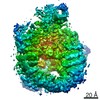
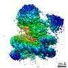
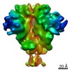
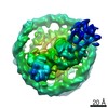
- PDB-4j9u: Crystal Structure of the TrkH/TrkA potassium transport complex -
+
Open data
ID or keywords:
Loading...
-
Basic information
Entry
Database: PDB / ID: 4j9u
Title
Crystal Structure of the TrkH/TrkA potassium transport complex
Components
Potassium uptake protein TrkA
Trk system potassium uptake protein TrkH
Keywords
TRANSPORT PROTEIN / RCK domain / potassium transport / membrane protein / Structural Genomics / PSI-Biology / New York Consortium on Membrane Protein Structure / NYCOMPS
Function / homology
Function and homology information
potassium:chloride symporter activity / potassium ion transmembrane transporter activity / potassium ion binding / potassium channel activity / potassium ion transmembrane transport / nucleotide binding / protein homodimerization activity / metal ion binding / identical protein binding / plasma membrane Similarity search - Function
Potassium uptake protein TrkA / TrkH potassium transport family / Cation transporter / Cation transport protein / Regulator of K+ conductance, C-terminal domain / : / TrkA-N domain / Regulator of K+ conductance, C-terminal / Regulator of K+ conductance, C-terminal domain superfamily / TrkA-C domain ...Potassium uptake protein TrkA / TrkH potassium transport family / Cation transporter / Cation transport protein / Regulator of K+ conductance, C-terminal domain / : / TrkA-N domain / Regulator of K+ conductance, C-terminal / Regulator of K+ conductance, C-terminal domain superfamily / TrkA-C domain / RCK C-terminal domain profile. / Regulator of K+ conductance, N-terminal / RCK N-terminal domain profile. / NAD(P)-binding Rossmann-like Domain / Alpha-Beta Plaits / NAD(P)-binding domain superfamily / Rossmann fold / 2-Layer Sandwich / 3-Layer(aba) Sandwich / Alpha Beta Similarity search - Domain/homology
: / NICOTINAMIDE-ADENINE-DINUCLEOTIDE / HEXATANTALUM DODECABROMIDE / Trk system potassium uptake protein TrkA / Trk system potassium uptake protein TrkH Similarity search - Component
Biological species
Vibrio parahaemolyticus (bacteria)
Method
X-RAY DIFFRACTION / SYNCHROTRON / SAD / Resolution: 3.8 Å
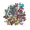
A: Trk system potassium uptake protein TrkH B: Trk system potassium uptake protein TrkH C: Trk system potassium uptake protein TrkH D: Trk system potassium uptake protein TrkH E: Potassium uptake protein TrkA F: Potassium uptake protein TrkA G: Potassium uptake protein TrkA H: Potassium uptake protein TrkA hetero molecules
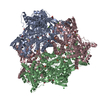
A: Trk system potassium uptake protein TrkH B: Trk system potassium uptake protein TrkH E: Potassium uptake protein TrkA F: Potassium uptake protein TrkA G: Potassium uptake protein TrkA H: Potassium uptake protein TrkA hetero molecules
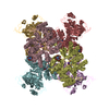
C: Trk system potassium uptake protein TrkH D: Trk system potassium uptake protein TrkH E: Potassium uptake protein TrkA F: Potassium uptake protein TrkA G: Potassium uptake protein TrkA H: Potassium uptake protein TrkA hetero molecules
 Movie
Movie Controller


 Open data
 Basic information
 Components
 Keywords
 Function and homology information
 Vibrio parahaemolyticus (bacteria)
 X-RAY DIFFRACTION /
  Authors
 Citation
 Structure visualization
 Downloads & links
 Other downloads


 PDBj
PDBj

 Assembly

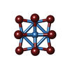
 Mass: 2044.535 Da / Num. of mol.: 18 / Source method: obtained synthetically / Formula: Br12Ta6
 Mass: 39.098 Da / Num. of mol.: 4 / Source method: obtained synthetically / Formula: K
 Mass: 663.425 Da / Num. of mol.: 4 / Source method: obtained synthetically / Formula: C21H27N7O14P2 / Comment: NAD*YM
 Sample preparation
 / Beamline: X25 / Wavelength: 1.2515 Å
 Processing