 Movie
Movie Controller

[English] 日本語
 Yorodumi
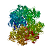
- PDB-6klo: Complex structure of Iota toxin enzymatic component (Ia) and bind... -
+ Open data

- Basic information
| Entry | Database: PDB / ID: 6klo | |||||||||
|---|---|---|---|---|---|---|---|---|---|---|
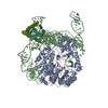
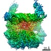
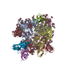
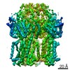
| Title | Complex structure of Iota toxin enzymatic component (Ia) and binding component (Ib) pore with short stem | |||||||||
 Components Components |
| |||||||||
 Keywords Keywords | TOXIN / Bacterial binary toxin / Protein translocation channel | |||||||||
| Function / homology |  Function and homology information Function and homology information | |||||||||
| Biological species |   Clostridium perfringens (bacteria) Clostridium perfringens (bacteria) | |||||||||
| Method | ELECTRON MICROSCOPY / single particle reconstruction / cryo EM / Resolution: 2.8 Å | |||||||||
 Authors Authors | Yoshida, T. / Yamada, T. / Kawamoto, A. / Mitsuoka, K. / Iwasaki, K. / Tsuge, H. | |||||||||
| Funding support |  Japan, 2items Japan, 2items
| |||||||||
 Citation Citation | Journal: Nat Struct Mol Biol / Year: 2020 Title: Cryo-EM structures reveal translocational unfolding in the clostridial binary iota toxin complex. Authors: Tomohito Yamada / Toru Yoshida / Akihiro Kawamoto / Kaoru Mitsuoka / Kenji Iwasaki / Hideaki Tsuge / Abstract: The iota toxin produced by Clostridium perfringens type E is a binary toxin comprising two independent polypeptides: Ia, an ADP-ribosyltransferase, and Ib, which is involved in cell binding and ...The iota toxin produced by Clostridium perfringens type E is a binary toxin comprising two independent polypeptides: Ia, an ADP-ribosyltransferase, and Ib, which is involved in cell binding and translocation of Ia across the cell membrane. Here we report cryo-EM structures of the translocation channel Ib-pore and its complex with Ia. The high-resolution Ib-pore structure demonstrates a similar structural framework to that of the catalytic ϕ-clamp of the anthrax protective antigen pore. However, the Ia-bound Ib-pore structure shows a unique binding mode of Ia: one Ia binds to the Ib-pore, and the Ia amino-terminal domain forms multiple weak interactions with two additional Ib-pore constriction sites. Furthermore, Ib-binding induces tilting and partial unfolding of the Ia N-terminal α-helix, permitting its extension to the ϕ-clamp gate. This new mechanism of N-terminal unfolding is crucial for protein translocation. | |||||||||
| History |
|
- Structure visualization
| Movie |
Movie viewer |
|---|---|
| Structure viewer | Molecule: MolmilJmol/JSmol |
- Downloads & links
-Download
| PDBx/mmCIF format | 6klo.cif.gz | 535.9 KB | Display | PDBx/mmCIF format |
|---|---|---|---|---|
| PDB format | pdb6klo.ent.gz | 427.3 KB | Display | PDB format |
| PDBx/mmJSON format | 6klo.json.gz | Tree view | PDBx/mmJSON format | |
| Others |  Other downloads Other downloads |
-Validation report
| Arichive directory | https://data.pdbj.org/pub/pdb/validation_reports/kl/6kloftp://data.pdbj.org/pub/pdb/validation_reports/kl/6klo | HTTPS FTP |
|---|
-Related structure data
| Related structure data |  0713MC  0720C  0721C  6klwC  6klxC M: map data used to model this data C: citing same article ( |
|---|---|
| Similar structure data |
-Links
 PDBj
PDBj
- Assembly
| Deposited unit | 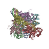
|
|---|---|
| 1 |
|
-Components
| #1: Protein | Mass: 74378.820 Da / Num. of mol.: 7 Source method: isolated from a genetically manipulated source Source: (gene. exp.) Clostridium perfringens (bacteria) / Plasmid: pGEX-4T-1 / Production host: #2: Protein | | Mass: 48061.988 Da / Num. of mol.: 1 Source method: isolated from a genetically manipulated source Source: (gene. exp.) Clostridium perfringens (bacteria) / Plasmid: pET15b / Production host: #3: Chemical | ChemComp-CA /   Mass: 40.078 Da / Num. of mol.: 14 / Source method: obtained synthetically / Formula: Ca Mass: 40.078 Da / Num. of mol.: 14 / Source method: obtained synthetically / Formula: CaHas ligand of interest | N | |
|---|
-Experimental details
-Experiment
| Experiment | Method: ELECTRON MICROSCOPY |
|---|---|
| EM experiment | Aggregation state: PARTICLE / 3D reconstruction method: single particle reconstruction |
- Sample preparation
| Component | Name: Complex structure of Iota toxin enzymatic component (Ia) and binding component (Ib) pore with short stem Type: COMPLEX / Entity ID: #1-#2 / Source: RECOMBINANT | ||||||||||||||||||||
|---|---|---|---|---|---|---|---|---|---|---|---|---|---|---|---|---|---|---|---|---|---|
| Molecular weight | Value: 0.57 MDa / Experimental value: NO | ||||||||||||||||||||
| Source (natural) | Organism: Clostridium perfringens (bacteria) | ||||||||||||||||||||
| Source (recombinant) | Organism: | ||||||||||||||||||||
| Buffer solution | pH: 7.5 | ||||||||||||||||||||
| Buffer component |
| ||||||||||||||||||||
| Specimen | Conc.: 0.48 mg/ml / Embedding applied: NO / Shadowing applied: NO / Staining applied: NO / Vitrification applied: YES | ||||||||||||||||||||
| Specimen support | Grid material: COPPER / Grid mesh size: 200 divisions/in. / Grid type: Quantifoil R1.2/1.3 | ||||||||||||||||||||
| Vitrification | Instrument: FEI VITROBOT MARK IV / Cryogen name: ETHANE / Humidity: 100 % / Chamber temperature: 277 K |
- Electron microscopy imaging
| Experimental equipment |  Model: Titan Krios / Image courtesy: FEI Company |
|---|---|
| Microscopy | Model: FEI TITAN KRIOS |
| Electron gun | Electron source:  FIELD EMISSION GUN / Accelerating voltage: 300 kV / Illumination mode: FLOOD BEAM FIELD EMISSION GUN / Accelerating voltage: 300 kV / Illumination mode: FLOOD BEAM |
| Electron lens | Mode: BRIGHT FIELD / Nominal magnification: 59000 X / Nominal defocus max: 2500 nm / Nominal defocus min: 800 nm / C2 aperture diameter: 50 µm |
| Specimen holder | Cryogen: NITROGEN / Specimen holder model: FEI TITAN KRIOS AUTOGRID HOLDER / Temperature (max): 78.9 K / Temperature (min): 78.9 K |
| Image recording | Average exposure time: 84.09 sec. / Electron dose: 50 e/Å2 / Detector mode: COUNTING / Film or detector model: FEI FALCON III (4k x 4k) / Num. of grids imaged: 1 / Num. of real images: 2151 |
| EM imaging optics | Spherical aberration corrector: Microscope was modified with a Cs corrector |
| Image scans | Width: 4096 / Height: 4096 |
- Processing
| Software | Name: PHENIX / Version: (1.15.2_3472: phenix.real_space_refine) / Classification: refinement | ||||||||||||||||||||||||||||||||||||||||
|---|---|---|---|---|---|---|---|---|---|---|---|---|---|---|---|---|---|---|---|---|---|---|---|---|---|---|---|---|---|---|---|---|---|---|---|---|---|---|---|---|---|
| EM software |
| ||||||||||||||||||||||||||||||||||||||||
| CTF correction | Type: PHASE FLIPPING AND AMPLITUDE CORRECTION | ||||||||||||||||||||||||||||||||||||||||
| Particle selection | Num. of particles selected: 871264 | ||||||||||||||||||||||||||||||||||||||||
| Symmetry | Point symmetry: C1 (asymmetric) | ||||||||||||||||||||||||||||||||||||||||
| 3D reconstruction | Resolution: 2.8 Å / Resolution method: FSC 0.143 CUT-OFF / Num. of particles: 135359 / Symmetry type: POINT | ||||||||||||||||||||||||||||||||||||||||
| Atomic model building | B value: 10 / Protocol: RIGID BODY FIT / Space: REAL | ||||||||||||||||||||||||||||||||||||||||
| Atomic model building | PDB-ID: 3J9C Accession code: 3J9C / Source name: PDB / Type: experimental model |

