Movie
Movie Controller

[English] 日本語
 Yorodumi
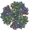
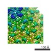
- PDB-6k33: Structure of PSI-isiA supercomplex from Thermosynechococcus vulcanus -
+ Open data

- Basic information
| Entry | Database: PDB / ID: 6k33 | |||||||||
|---|---|---|---|---|---|---|---|---|---|---|
| Title | Structure of PSI-isiA supercomplex from Thermosynechococcus vulcanus | |||||||||
 Components Components |
| |||||||||
 Keywords Keywords | PHOTOSYNTHESIS / Electron Transport | |||||||||
| Function / homology |  Function and homology information Function and homology informationphotosystem I reaction center / photosystem I / photosystem I / photosynthetic electron transport in photosystem I / chlorophyll binding / plasma membrane-derived thylakoid membrane / photosynthesis / 4 iron, 4 sulfur cluster binding / electron transfer activity / oxidoreductase activity ...photosystem I reaction center / photosystem I / photosystem I / photosynthetic electron transport in photosystem I / chlorophyll binding / plasma membrane-derived thylakoid membrane / photosynthesis / 4 iron, 4 sulfur cluster binding / electron transfer activity / oxidoreductase activity / magnesium ion binding / metal ion binding Similarity search - Function | |||||||||
| Biological species |  Thermosynechococcus vulcanus (bacteria) Thermosynechococcus vulcanus (bacteria) | |||||||||
| Method | ELECTRON MICROSCOPY / single particle reconstruction / cryo EM / Resolution: 2.74 Å | |||||||||
 Authors Authors | Akita, F. / Nagao, R. / Kato, K. / Shen, J.R. / Miyazaki, N. | |||||||||
 Citation Citation | Journal: Commun Biol / Year: 2020 Title: Structure of a cyanobacterial photosystem I surrounded by octadecameric IsiA antenna proteins. Authors: Fusamichi Akita / Ryo Nagao / Koji Kato / Yoshiki Nakajima / Makio Yokono / Yoshifumi Ueno / Takehiro Suzuki / Naoshi Dohmae / Jian-Ren Shen / Seiji Akimoto / Naoyuki Miyazaki /  Abstract: Iron-stress induced protein A (IsiA) is a chlorophyll-binding membrane-spanning protein in photosynthetic prokaryote cyanobacteria, and is associated with photosystem I (PSI) trimer cores, but its ...Iron-stress induced protein A (IsiA) is a chlorophyll-binding membrane-spanning protein in photosynthetic prokaryote cyanobacteria, and is associated with photosystem I (PSI) trimer cores, but its structural and functional significance in light harvesting remains unclear. Here we report a 2.7-Å resolution cryo-electron microscopic structure of a supercomplex between PSI core trimer and IsiA from a thermophilic cyanobacterium Thermosynechococcus vulcanus. The structure showed that 18 IsiA subunits form a closed ring surrounding a PSI trimer core. Detailed arrangement of pigments within the supercomplex, as well as molecular interactions between PSI and IsiA and among IsiAs, were resolved. Time-resolved fluorescence spectra of the PSI-IsiA supercomplex showed clear excitation-energy transfer from IsiA to PSI, strongly indicating that IsiA functions as an energy donor, but not an energy quencher, in the supercomplex. These structural and spectroscopic findings provide important insights into the excitation-energy-transfer and subunit assembly mechanisms in the PSI-IsiA supercomplex. | |||||||||
| History |
|
- Structure visualization
| Movie |
Movie viewer |
|---|---|
| Structure viewer | Molecule: MolmilJmol/JSmol |
- Downloads & links
-Download
| PDBx/mmCIF format | 6k33.cif.gz | 2.8 MB | Display | PDBx/mmCIF format |
|---|---|---|---|---|
| PDB format | pdb6k33.ent.gz | Display | PDB format | |
| PDBx/mmJSON format | 6k33.json.gz | Tree view | PDBx/mmJSON format | |
| Others |  Other downloads Other downloads |
-Validation report
| Summary document | 6k33_validation.pdf.gz | 37.6 MB | Display | wwPDB validaton report |
|---|---|---|---|---|
| Full document | 6k33_full_validation.pdf.gz | 40.3 MB | Display | |
| Data in XML | 6k33_validation.xml.gz | 627.2 KB | Display | |
| Data in CIF | 6k33_validation.cif.gz | 773.7 KB | Display | |
| Arichive directory | https://data.pdbj.org/pub/pdb/validation_reports/k3/6k33ftp://data.pdbj.org/pub/pdb/validation_reports/k3/6k33 | HTTPS FTP |
-Related structure data
| Related structure data | 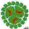 9908MC M: map data used to model this data C: citing same article ( |
|---|---|
| Similar structure data |


-Links
 PDBj
PDBj


- Assembly
| Deposited unit | 
|
|---|---|
| 1 |
|
-Components
-Photosystem I P700 chlorophyll a apoprotein ... , 2 types, 6 molecules aAbAcAaBbBcB
| #1: Protein | Mass: 83283.773 Da / Num. of mol.: 3 / Source method: isolated from a natural source / Source: (natural) Thermosynechococcus vulcanus (bacteria) / References: UniProt: P25936, photosystem I#2: Protein | Mass: 82992.453 Da / Num. of mol.: 3 / Source method: isolated from a natural source / Source: (natural) Thermosynechococcus vulcanus (bacteria) / References: UniProt: P0A409, photosystem I |
|---|
-Protein , 2 types, 21 molecules aCbCcCa1a2a3a4a5a6b1b2b3b4b5b6c1c2c3c4c5c6
| #3: Protein | Mass: 8678.011 Da / Num. of mol.: 3 / Source method: isolated from a natural source / Source: (natural) Thermosynechococcus vulcanus (bacteria) / References: UniProt: P0A417, photosystem I#13: Protein | Mass: 39284.332 Da / Num. of mol.: 18 / Source method: isolated from a natural source / Source: (natural) Thermosynechococcus vulcanus (bacteria) |
|---|
-Photosystem I reaction center subunit ... , 9 types, 27 molecules aDbDcDaEbEcEaFbFcFaIbIcIaJbJcJaKbKcKaLbLcLaMbMcMaXbXcX
| #4: Protein | Mass: 15258.297 Da / Num. of mol.: 3 / Source method: isolated from a natural source / Source: (natural) Thermosynechococcus vulcanus (bacteria) / References: UniProt: P0A422#5: Protein | Mass: 8268.290 Da / Num. of mol.: 3 / Source method: isolated from a natural source / Source: (natural) Thermosynechococcus vulcanus (bacteria)#6: Protein | Mass: 17716.586 Da / Num. of mol.: 3 / Source method: isolated from a natural source / Source: (natural) Thermosynechococcus vulcanus (bacteria)#7: Protein/peptide | Mass: 4297.234 Da / Num. of mol.: 3 / Source method: isolated from a natural source / Source: (natural) Thermosynechococcus vulcanus (bacteria)#8: Protein/peptide | Mass: 4770.698 Da / Num. of mol.: 3 / Source method: isolated from a natural source / Source: (natural) Thermosynechococcus vulcanus (bacteria)#9: Protein | Mass: 8668.153 Da / Num. of mol.: 3 / Source method: isolated from a natural source / Source: (natural) Thermosynechococcus vulcanus (bacteria) / References: UniProt: P23318#10: Protein | Mass: 16156.569 Da / Num. of mol.: 3 / Source method: isolated from a natural source / Source: (natural) Thermosynechococcus vulcanus (bacteria)#11: Protein/peptide | Mass: 3426.115 Da / Num. of mol.: 3 / Source method: isolated from a natural source / Source: (natural) Thermosynechococcus vulcanus (bacteria)#12: Protein/peptide | Mass: 3845.508 Da / Num. of mol.: 3 / Source method: isolated from a natural source / Source: (natural) Thermosynechococcus vulcanus (bacteria) |
|---|
-Non-polymers , 9 types, 780 molecules 


| #14: Chemical |  Mass: 893.489 Da / Num. of mol.: 3 / Source method: obtained synthetically / Formula: C55H72MgN4O5 Mass: 893.489 Da / Num. of mol.: 3 / Source method: obtained synthetically / Formula: C55H72MgN4O5#15: Chemical | ChemComp-CLA /  Mass: 893.489 Da / Num. of mol.: 588 / Source method: obtained synthetically / Formula: C55H72MgN4O5 Mass: 893.489 Da / Num. of mol.: 588 / Source method: obtained synthetically / Formula: C55H72MgN4O5#16: Chemical | ChemComp-PQN /  Mass: 450.696 Da / Num. of mol.: 6 / Source method: obtained synthetically / Formula: C31H46O2 Mass: 450.696 Da / Num. of mol.: 6 / Source method: obtained synthetically / Formula: C31H46O2#17: Chemical | ChemComp-SF4 /  Mass: 351.640 Da / Num. of mol.: 9 / Source method: obtained synthetically / Formula: Fe4S4 Mass: 351.640 Da / Num. of mol.: 9 / Source method: obtained synthetically / Formula: Fe4S4#18: Chemical | ChemComp-BCR /  Mass: 536.873 Da / Num. of mol.: 138 / Source method: obtained synthetically / Formula: C40H56 Mass: 536.873 Da / Num. of mol.: 138 / Source method: obtained synthetically / Formula: C40H56#19: Chemical | ChemComp-LHG /  Mass: 722.970 Da / Num. of mol.: 9 / Source method: obtained synthetically / Formula: C38H75O10P / Comment: phospholipid*YM Mass: 722.970 Da / Num. of mol.: 9 / Source method: obtained synthetically / Formula: C38H75O10P / Comment: phospholipid*YM#20: Chemical |  Mass: 787.158 Da / Num. of mol.: 3 / Source method: obtained synthetically / Formula: C45H86O10 Mass: 787.158 Da / Num. of mol.: 3 / Source method: obtained synthetically / Formula: C45H86O10#21: Chemical |  Mass: 40.078 Da / Num. of mol.: 3 / Source method: obtained synthetically / Formula: Ca Mass: 40.078 Da / Num. of mol.: 3 / Source method: obtained synthetically / Formula: Ca#22: Water | ChemComp-HOH / | Mass: 18.015 Da / Num. of mol.: 21 / Source method: isolated from a natural source / Formula: H2O |
|---|
-Details
| Has ligand of interest | N |
|---|---|
| Has protein modification | Y |
-Experimental details
-Experiment
| Experiment | Method: ELECTRON MICROSCOPY |
|---|---|
| EM experiment | Aggregation state: PARTICLE / 3D reconstruction method: single particle reconstruction |
- Sample preparation
| Component | Name: PSI-isiA / Type: COMPLEX / Entity ID: #1-#13 / Source: NATURAL | |||||||||||||||
|---|---|---|---|---|---|---|---|---|---|---|---|---|---|---|---|---|
| Molecular weight | Value: 1.9 MDa / Experimental value: NO | |||||||||||||||
| Source (natural) | Organism: Thermosynechococcus vulcanus (bacteria) | |||||||||||||||
| Buffer solution | pH: 7 | |||||||||||||||
| Buffer component |
| |||||||||||||||
| Specimen | Conc.: 2 mg/ml / Embedding applied: NO / Shadowing applied: NO / Staining applied: NO / Vitrification applied: YES | |||||||||||||||
| Specimen support | Grid material: COPPER / Grid mesh size: 300 divisions/in. / Grid type: Quantifoil R2/1 | |||||||||||||||
| Vitrification | Instrument: FEI VITROBOT MARK IV / Cryogen name: ETHANE / Humidity: 100 % / Chamber temperature: 277 K |
- Electron microscopy imaging
| Experimental equipment |  Model: Titan Krios / Image courtesy: FEI Company |
|---|---|
| Microscopy | Model: FEI TITAN KRIOS |
| Electron gun | Electron source:  FIELD EMISSION GUN / Accelerating voltage: 300 kV / Illumination mode: FLOOD BEAM FIELD EMISSION GUN / Accelerating voltage: 300 kV / Illumination mode: FLOOD BEAM |
| Electron lens | Mode: BRIGHT FIELD |
| Image recording | Electron dose: 40 e/Å2 / Film or detector model: FEI FALCON III (4k x 4k) |
- Processing
| EM software |
| ||||||||||||||||||||||||||||||||||||
|---|---|---|---|---|---|---|---|---|---|---|---|---|---|---|---|---|---|---|---|---|---|---|---|---|---|---|---|---|---|---|---|---|---|---|---|---|---|
| CTF correction | Type: PHASE FLIPPING AND AMPLITUDE CORRECTION | ||||||||||||||||||||||||||||||||||||
| Symmetry | Point symmetry: C3 (3 fold cyclic) | ||||||||||||||||||||||||||||||||||||
| 3D reconstruction | Resolution: 2.74 Å / Resolution method: FSC 0.143 CUT-OFF / Num. of particles: 303983 / Algorithm: FOURIER SPACE / Num. of class averages: 1 / Symmetry type: POINT | ||||||||||||||||||||||||||||||||||||
| Atomic model building | Protocol: FLEXIBLE FIT / Space: REAL / Target criteria: Correlation coefficient | ||||||||||||||||||||||||||||||||||||
| Atomic model building | PDB-ID: 1JB0 Accession code: 1JB0 / Source name: PDB / Type: experimental model |