 Movie
Movie Controller


+ Open data

- Basic information
| Entry | Database: PDB / ID: 6k1h | ||||||||||||||||||||||||||||||||||||||||||||||||||||||||||||||||||
|---|---|---|---|---|---|---|---|---|---|---|---|---|---|---|---|---|---|---|---|---|---|---|---|---|---|---|---|---|---|---|---|---|---|---|---|---|---|---|---|---|---|---|---|---|---|---|---|---|---|---|---|---|---|---|---|---|---|---|---|---|---|---|---|---|---|---|---|
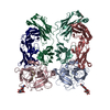
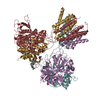
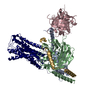
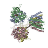
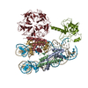
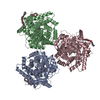
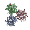
| Title | Structure of membrane protein | ||||||||||||||||||||||||||||||||||||||||||||||||||||||||||||||||||
 Components Components |
| ||||||||||||||||||||||||||||||||||||||||||||||||||||||||||||||||||
 Keywords Keywords | PROTEIN TRANSPORT / PTS / ManYZ / transporter / Mannose | ||||||||||||||||||||||||||||||||||||||||||||||||||||||||||||||||||
| Function / homology |  Function and homology information Function and homology informationmannose transmembrane transport / protein-N(PI)-phosphohistidine-mannose phosphotransferase system transporter activity / fructose import across plasma membrane / N-acetylglucosamine transport / D-glucose import across plasma membrane / phosphoenolpyruvate-dependent sugar phosphotransferase system / transmembrane transporter complex / membrane / plasma membrane Similarity search - Function | ||||||||||||||||||||||||||||||||||||||||||||||||||||||||||||||||||
| Biological species |  | ||||||||||||||||||||||||||||||||||||||||||||||||||||||||||||||||||
| Method | ELECTRON MICROSCOPY / single particle reconstruction / cryo EM / Resolution: 3.52 Å | ||||||||||||||||||||||||||||||||||||||||||||||||||||||||||||||||||
 Authors Authors | Wang, J.W. / Zeng, J.W. | ||||||||||||||||||||||||||||||||||||||||||||||||||||||||||||||||||
| Funding support |  China, 3items China, 3items
| ||||||||||||||||||||||||||||||||||||||||||||||||||||||||||||||||||
 Citation Citation | Journal: Cell Res / Year: 2019 Title: Structure of the mannose transporter of the bacterial phosphotransferase system. Authors: Xueli Liu / Jianwei Zeng / Kai Huang / Jiawei Wang / | ||||||||||||||||||||||||||||||||||||||||||||||||||||||||||||||||||
| History |
|
- Structure visualization
| Movie |
Movie viewer |
|---|---|
| Structure viewer | Molecule: MolmilJmol/JSmol |
- Downloads & links
-Download
| PDBx/mmCIF format | 6k1h.cif.gz | 264.6 KB | Display | PDBx/mmCIF format |
|---|---|---|---|---|
| PDB format | pdb6k1h.ent.gz | 214.4 KB | Display | PDB format |
| PDBx/mmJSON format | 6k1h.json.gz | Tree view | PDBx/mmJSON format | |
| Others |  Other downloads Other downloads |
-Validation report
| Arichive directory | https://data.pdbj.org/pub/pdb/validation_reports/k1/6k1hftp://data.pdbj.org/pub/pdb/validation_reports/k1/6k1h | HTTPS FTP |
|---|
-Related structure data
| Related structure data |  9906MC M: map data used to model this data C: citing same article ( |
|---|---|
| Similar structure data |

-Links
 PDBj
PDBj


- Assembly
| Deposited unit | 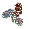
|
|---|---|
| 1 |
|
-Components
| #1: Protein | Mass: 30983.373 Da / Num. of mol.: 3 Source method: isolated from a genetically manipulated source Source: (gene. exp.) Gene: D8B36_09735 / Variant: K-12 Production host: Variant (production host): BL21 / References: UniProt: A0A387CW08, UniProt: P69805*PLUS #2: Protein | Mass: 28752.783 Da / Num. of mol.: 3 Source method: isolated from a genetically manipulated source Source: (gene. exp.) Strain: K12 / Gene: manY, pel, ptsP, b1818, JW1807 / Variant: K-12 Production host: Variant (production host): BL21 / References: UniProt: P69801 #3: Sugar |   Type: D-saccharide, alpha linking / Mass: 180.156 Da / Num. of mol.: 3 Type: D-saccharide, alpha linking / Mass: 180.156 Da / Num. of mol.: 3Source method: isolated from a genetically manipulated source Formula: C6H12O6 / Feature type: SUBJECT OF INVESTIGATION Has ligand of interest | Y | Has protein modification | N | |
|---|
-Experimental details
-Experiment
| Experiment | Method: ELECTRON MICROSCOPY |
|---|---|
| EM experiment | Aggregation state: PARTICLE / 3D reconstruction method: single particle reconstruction |
- Sample preparation
| Component | Name: ManYZ trimer complex / Type: COMPLEX / Entity ID: #1-#2 / Source: RECOMBINANT |
|---|---|
| Molecular weight | Value: 176 kDa/nm / Experimental value: YES |
| Source (natural) | Organism: |
| Source (recombinant) | Organism: Strain: BL21 |
| Buffer solution | pH: 8 |
| Specimen | Embedding applied: NO / Shadowing applied: NO / Staining applied: NO / Vitrification applied: YES |
| Vitrification | Instrument: FEI VITROBOT MARK IV / Cryogen name: ETHANE / Humidity: 100 % / Chamber temperature: 281 K |
- Electron microscopy imaging
| Experimental equipment |  Model: Titan Krios / Image courtesy: FEI Company |
|---|---|
| Microscopy | Model: FEI TITAN KRIOS |
| Electron gun | Electron source:  FIELD EMISSION GUN / Accelerating voltage: 300 kV / Illumination mode: FLOOD BEAM FIELD EMISSION GUN / Accelerating voltage: 300 kV / Illumination mode: FLOOD BEAM |
| Electron lens | Mode: BRIGHT FIELD |
| Image recording | Electron dose: 50 e/Å2 / Detector mode: SUPER-RESOLUTION / Film or detector model: GATAN K2 SUMMIT (4k x 4k) |
- Processing
| Software | Name: PHENIX / Version: 1.18.1_3865: / Classification: refinement | ||||||||||||||||||||||||
|---|---|---|---|---|---|---|---|---|---|---|---|---|---|---|---|---|---|---|---|---|---|---|---|---|---|
| EM software | Name: PHENIX / Category: model refinement | ||||||||||||||||||||||||
| CTF correction | Type: PHASE FLIPPING AND AMPLITUDE CORRECTION | ||||||||||||||||||||||||
| 3D reconstruction | Resolution: 3.52 Å / Resolution method: FSC 0.143 CUT-OFF / Num. of particles: 309996 / Symmetry type: POINT | ||||||||||||||||||||||||
| Refine LS restraints |
|
