[English] 日本語
Yorodumi
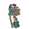
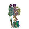
- PDB-6j5j: Cryo-EM structure of the mammalian E-state ATP synthase -

+
Open data


ID or keywords:

Loading...

-
Basic information

Entry
Database: PDB / ID: 6j5j
TitleCryo-EM structure of the mammalian E-state ATP synthase
Components
  • (ATP synthase ...) x 15
  • ATP synthase-coupling factor 6, mitochondrial
  • ATPase inhibitor, mitochondrial
  • Mitochondrial H+ transporting ATP synthase subunit c isoform 1
  • subunit k analog
KeywordsMEMBRANE PROTEIN
Function / homology
Function and homology information


Formation of ATP by chemiosmotic coupling / Cristae formation / negative regulation of mitochondrial ATP synthesis coupled proton transport / Mitochondrial protein import / angiostatin binding / ATPase inhibitor activity / Mitochondrial protein degradation / negative regulation of hydrolase activity / : / : ...Formation of ATP by chemiosmotic coupling / Cristae formation / negative regulation of mitochondrial ATP synthesis coupled proton transport / Mitochondrial protein import / angiostatin binding / ATPase inhibitor activity / Mitochondrial protein degradation / negative regulation of hydrolase activity / : / : / proton-transporting ATP synthase complex / heme biosynthetic process / : / : / : / proton-transporting ATP synthase complex, coupling factor F(o) / negative regulation of endothelial cell proliferation / proton motive force-driven ATP synthesis / proton motive force-driven mitochondrial ATP synthesis / proton transmembrane transporter activity / proton-transporting ATP synthase complex, catalytic core F(1) / H+-transporting two-sector ATPase / proton-transporting ATPase activity, rotational mechanism / proton-transporting ATP synthase activity, rotational mechanism / proton transmembrane transport / erythrocyte differentiation / ADP binding / ATPase binding / mitochondrial inner membrane / calmodulin binding / lipid binding / cell surface / ATP hydrolysis activity / protein-containing complex / mitochondrion / ATP binding / identical protein binding / plasma membrane
Similarity search - Function
ATP synthase, F1 complex, epsilon subunit superfamily, mitochondrial / ATP synthase delta/epsilon subunit, C-terminal domain / ATP Synthase; domain 1 / F0F1 ATP synthase delta/epsilon subunit, N-terminal / ATP synthase, F1 complex, gamma subunit / Atp Synthase Epsilon Chain; Chain: I; / ATP synthase membrane subunit K / ATP synthase regulation / ATP synthase alpha/beta chain, C-terminal domain / F1F0 ATP synthase subunit C ...ATP synthase, F1 complex, epsilon subunit superfamily, mitochondrial / ATP synthase delta/epsilon subunit, C-terminal domain / ATP Synthase; domain 1 / F0F1 ATP synthase delta/epsilon subunit, N-terminal / ATP synthase, F1 complex, gamma subunit / Atp Synthase Epsilon Chain; Chain: I; / ATP synthase membrane subunit K / ATP synthase regulation / ATP synthase alpha/beta chain, C-terminal domain / F1F0 ATP synthase subunit C / F1FO ATP Synthase / Lysin / Thrombin, subunit H - #170 / ATP synthase protein 8, metazoa / Mitochondrial ATPase inhibitor / Mitochondrial F1-F0 ATP synthase subunit F, predicted / ATP synthase protein 8, mammals / ATP synthase protein 8 / Mitochondrial ATPase inhibitor, IATP / Mitochondrial F1F0-ATP synthase, subunit f / Elongation Factor Tu (Ef-tu); domain 3 - #20 / ATP synthase-coupling factor 6, mitochondrial / ATP synthase-coupling factor 6 superfamily, mitochondrial / Mitochondrial ATP synthase coupling factor 6 / Pyruvate Kinase; Chain: A, domain 1 / Bovine Mitochondrial F1-ATPase, ATP Synthase Beta Chain; Chain D, domain3 / Bovine Mitochondrial F1-atpase; Atp Synthase Beta Chain; Chain D, domain 3 / : / Metazoan delta subunit of F1F0-ATP synthase, C-terminal domain / ATP synthase, gamma subunit, helix hairpin domain / ATP synthase delta/epsilon subunit, C-terminal domain superfamily / ATP synthase, F0 complex, subunit B/MI25 / ATP synthase, F0 complex, subunit B / Mitochondrial ATP synthase B chain precursor (ATP-synt_B) / ATP synthase, F0 complex, subunit D, mitochondrial / ATP synthase D chain, mitochondrial (ATP5H) / ATP synthase, F0 complex, subunit D superfamily, mitochondrial / ATP synthase, F0 complex, subunit A, bacterial/mitochondria / ATP synthase, F1 complex, epsilon subunit, mitochondrial / ATP synthase, F1 complex, epsilon subunit superfamily, mitochondrial / Mitochondrial ATP synthase epsilon chain / ATPase, OSCP/delta subunit, conserved site / ATP synthase delta (OSCP) subunit signature. / F1F0 ATP synthase OSCP/delta subunit, N-terminal domain superfamily / ATP synthase, F0 complex, subunit A / ATP synthase, F0 complex, subunit A, active site / ATP synthase, F0 complex, subunit A superfamily / ATP synthase A chain / ATP synthase a subunit signature. / ATPase, OSCP/delta subunit / ATP synthase delta (OSCP) subunit / ATP synthase, F1 complex, delta/epsilon subunit / ATP synthase, F1 complex, delta/epsilon subunit, N-terminal / F0F1 ATP synthase delta/epsilon subunit, N-terminal / ATP synthase, Delta/Epsilon chain, beta-sandwich domain / ATP synthase, F0 complex, subunit C / F1F0 ATP synthase subunit C superfamily / ATP synthase, F0 complex, subunit C, DCCD-binding site / ATP synthase c subunit signature. / ATP synthase, F1 complex, gamma subunit conserved site / ATP synthase gamma subunit signature. / ATP synthase, F1 complex, beta subunit / ATP synthase, alpha subunit, C-terminal domain superfamily / : / ATP synthase, F1 complex, gamma subunit / ATP synthase, F1 complex, gamma subunit superfamily / ATP synthase / ATP synthase, alpha subunit, C-terminal / ATP synthase, F1 complex, alpha subunit / ATP synthase, F1 complex, alpha subunit nucleotide-binding domain / ATP synthase alpha/beta chain, C terminal domain / V-ATPase proteolipid subunit C-like domain / F/V-ATP synthase subunit C superfamily / ATP synthase subunit C / ATPase, F1/V1 complex, beta/alpha subunit, C-terminal / C-terminal domain of V and A type ATP synthase / ATP synthase subunit alpha, N-terminal domain-like superfamily / ATPase, F1/V1/A1 complex, alpha/beta subunit, N-terminal domain superfamily / ATPase, F1/V1/A1 complex, alpha/beta subunit, N-terminal domain / ATP synthase alpha/beta family, beta-barrel domain / ATPase, alpha/beta subunit, nucleotide-binding domain, active site / ATP synthase alpha and beta subunits signature. / Elongation Factor Tu (Ef-tu); domain 3 / ATPase, F1/V1/A1 complex, alpha/beta subunit, nucleotide-binding domain / ATP synthase alpha/beta family, nucleotide-binding domain / Single alpha-helices involved in coiled-coils or other helix-helix interfaces / Helix Hairpins / Thrombin, subunit H / P-loop containing nucleotide triphosphate hydrolases / ATPases associated with a variety of cellular activities / AAA+ ATPase domain / Up-down Bundle / Beta Barrel / Sandwich / Rossmann fold / P-loop containing nucleoside triphosphate hydrolase / Orthogonal Bundle / 3-Layer(aba) Sandwich / Mainly Beta / Mainly Alpha
Similarity search - Domain/homology
ADENOSINE-5'-DIPHOSPHATE / ADENOSINE-5'-TRIPHOSPHATE / ATP synthase F1 subunit delta / ATP synthase subunit b / ATP synthase subunit gamma / ATP synthase subunit d, mitochondrial / ATP synthase, H+ transporting, mitochondrial F1 complex, epsilon subunit / ATP synthase membrane subunit K, mitochondrial / ATP synthase subunit beta / ATP synthase-coupling factor 6, mitochondrial ...ADENOSINE-5'-DIPHOSPHATE / ADENOSINE-5'-TRIPHOSPHATE / ATP synthase F1 subunit delta / ATP synthase subunit b / ATP synthase subunit gamma / ATP synthase subunit d, mitochondrial / ATP synthase, H+ transporting, mitochondrial F1 complex, epsilon subunit / ATP synthase membrane subunit K, mitochondrial / ATP synthase subunit beta / ATP synthase-coupling factor 6, mitochondrial / ATP synthase subunit alpha, mitochondrial / ATPase inhibitor, mitochondrial / ATP synthase subunit O, mitochondrial / ATP synthase protein 8 / ATP synthase subunit a / ATP synthase lipid-binding protein / ATP synthase subunit f, mitochondrial
Similarity search - Component
Biological speciesSus scrofa (pig)
MethodELECTRON MICROSCOPY / single particle reconstruction / cryo EM / Resolution: 3.45 Å
AuthorsGu, J. / Zhang, L. / Yi, J. / Yang, M.
Funding support China, 3items
OrganizationGrant numberCountry
Ministry of Science and Technology (China)2017YFA0504600 and 2016YFA0501100 China
National Natural Science Foundation of China31625008 China
National Natural Science Foundation of China21532004, 31570733 and 31800620 China
CitationJournal: Science / Year: 2019
Title: Cryo-EM structure of the mammalian ATP synthase tetramer bound with inhibitory protein IF1.
Authors: Jinke Gu / Laixing Zhang / Shuai Zong / Runyu Guo / Tianya Liu / Jingbo Yi / Peiyi Wang / Wei Zhuo / Maojun Yang /
Abstract: The mitochondrial adenosine triphosphate (ATP) synthase produces most of the ATP required by mammalian cells. We isolated porcine tetrameric ATP synthase and solved its structure at 6.2-angstrom ...The mitochondrial adenosine triphosphate (ATP) synthase produces most of the ATP required by mammalian cells. We isolated porcine tetrameric ATP synthase and solved its structure at 6.2-angstrom resolution using a single-particle cryo-electron microscopy method. Two classical V-shaped ATP synthase dimers lie antiparallel to each other to form an H-shaped ATP synthase tetramer, as viewed from the matrix. ATP synthase inhibitory factor subunit 1 (IF1) is a well-known in vivo inhibitor of mammalian ATP synthase at low pH. Two IF1 dimers link two ATP synthase dimers, which is consistent with the ATP synthase tetramer adopting an inhibited state. Within the tetramer, we refined structures of intact ATP synthase in two different rotational conformations at 3.34- and 3.45-Å resolution.
History
DepositionJan 11, 2019Deposition site: PDBJ / Processing site: PDBJ
Revision 1.0Jun 26, 2019Provider: repository / Type: Initial release
Revision 1.1Nov 6, 2019Group: Data collection / Other / Category: cell / Item: _cell.Z_PDB
Revision 1.2Mar 27, 2024Group: Data collection / Database references / Derived calculations
Category: chem_comp_atom / chem_comp_bond ...chem_comp_atom / chem_comp_bond / database_2 / struct_conn
Item: _database_2.pdbx_DOI / _database_2.pdbx_database_accession ..._database_2.pdbx_DOI / _database_2.pdbx_database_accession / _struct_conn.pdbx_dist_value / _struct_conn.ptnr1_auth_asym_id / _struct_conn.ptnr1_auth_comp_id / _struct_conn.ptnr1_auth_seq_id / _struct_conn.ptnr1_label_asym_id / _struct_conn.ptnr1_label_atom_id / _struct_conn.ptnr1_label_comp_id / _struct_conn.ptnr1_label_seq_id / _struct_conn.ptnr2_auth_asym_id / _struct_conn.ptnr2_auth_seq_id / _struct_conn.ptnr2_label_asym_id

-
Structure visualization

Movie
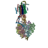
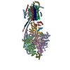
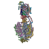
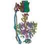
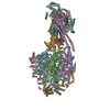
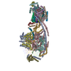
  • Deposited structure unit
  • Imaged by Jmol
  • Download
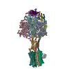
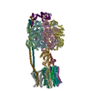
  • Superimposition on EM map
  • EMDB-0669
  • Imaged by UCSF Chimera
  • Download
Movie viewer
Structure viewerMolecule:
MolmilJmol/JSmol

Downloads & links

-
Assembly

Deposited unit
A: ATP synthase subunit alpha, mitochondrial
B: ATP synthase subunit alpha, mitochondrial
C: ATP synthase subunit alpha, mitochondrial
D: ATP synthase subunit beta
E: ATP synthase subunit beta
F: ATP synthase subunit beta
J: ATPase inhibitor, mitochondrial
G: ATP synthase subunit gamma
H: ATP synthase subunit delta, mitochondrial
I: ATP synthase F1 subunit epsilon
S: ATP synthase subunit O, mitochondrial
b: ATP synthase peripheral stalk-membrane subunit b
c: ATP synthase-coupling factor 6, mitochondrial
d: ATP synthase subunit d, mitochondrial
e: ATP synthase subunit e
f: ATP synthase subunit f, mitochondrial
g: ATP synthase subunit g
i: ATP synthase membrane subunit DAPIT
k: subunit k analog
8: ATP synthase protein 8
a: ATP synthase subunit a
K: Mitochondrial H+ transporting ATP synthase subunit c isoform 1
L: Mitochondrial H+ transporting ATP synthase subunit c isoform 1
M: Mitochondrial H+ transporting ATP synthase subunit c isoform 1
N: Mitochondrial H+ transporting ATP synthase subunit c isoform 1
O: Mitochondrial H+ transporting ATP synthase subunit c isoform 1
P: Mitochondrial H+ transporting ATP synthase subunit c isoform 1
Q: Mitochondrial H+ transporting ATP synthase subunit c isoform 1
R: Mitochondrial H+ transporting ATP synthase subunit c isoform 1
u: ATP synthase membrane subunit 6.8PL
hetero molecules


Theoretical massNumber of molelcules
Total (without water)573,57740
Polymers571,07930
Non-polymers2,49710
Water00
1


  • Idetical with deposited unit
  • defined by author
  • Evidence: native gel electrophoresis
TypeNameSymmetry operationNumber
identity operation1_5551

-
Components

-
ATP synthase ... , 15 types, 19 molecules ABCDEFGHISbdefgi8au

#1: Protein ATP synthase subunit alpha, mitochondrial / ATP synthase F1 subunit alpha / ATP synthase subunit alpha heart isoform / mitochondrial / ATP ...ATP synthase F1 subunit alpha / ATP synthase subunit alpha heart isoform / mitochondrial / ATP synthase subunit alpha liver isoform


Mass: 55171.105 Da / Num. of mol.: 3 / Source method: isolated from a natural source / Source: (natural) Sus scrofa (pig) / References: UniProt: P80021
#2: Protein ATP synthase subunit beta


Mass: 50606.652 Da / Num. of mol.: 3 / Source method: isolated from a natural source / Source: (natural) Sus scrofa (pig)
References: UniProt: K7GLT8, H+-transporting two-sector ATPase
#4: Protein ATP synthase subunit gamma


Mass: 30121.650 Da / Num. of mol.: 1 / Source method: isolated from a natural source / Source: (natural) Sus scrofa (pig) / References: UniProt: A0A287A9I8
#5: Protein ATP synthase subunit delta, mitochondrial


Mass: 13852.506 Da / Num. of mol.: 1 / Source method: isolated from a natural source / Source: (natural) Sus scrofa (pig) / References: UniProt: A0A286ZYL7
#6: Protein/peptide ATP synthase F1 subunit epsilon / ATP synthase / epsilon subunit


Mass: 5371.264 Da / Num. of mol.: 1 / Source method: isolated from a natural source / Source: (natural) Sus scrofa (pig) / References: UniProt: A5GFX4
#7: Protein ATP synthase subunit O, mitochondrial / ATP synthase peripheral stalk subunit OSCP


Mass: 20561.279 Da / Num. of mol.: 1 / Source method: isolated from a natural source / Source: (natural) Sus scrofa (pig) / References: UniProt: Q2EN81
#8: Protein ATP synthase peripheral stalk-membrane subunit b


Mass: 24002.934 Da / Num. of mol.: 1 / Source method: isolated from a natural source / Source: (natural) Sus scrofa (pig) / References: UniProt: A0A286ZYM6
#10: Protein ATP synthase subunit d, mitochondrial


Mass: 16904.473 Da / Num. of mol.: 1 / Source method: isolated from a natural source / Source: (natural) Sus scrofa (pig) / References: UniProt: A0A287B4I0
#11: Protein ATP synthase subunit e


Mass: 5379.623 Da / Num. of mol.: 1 / Source method: isolated from a natural source / Source: (natural) Sus scrofa (pig)
#12: Protein ATP synthase subunit f, mitochondrial / ATP synthase membrane subunit f


Mass: 10197.959 Da / Num. of mol.: 1 / Source method: isolated from a natural source / Source: (natural) Sus scrofa (pig) / References: UniProt: Q95339
#13: Protein ATP synthase subunit g


Mass: 7166.825 Da / Num. of mol.: 1 / Source method: isolated from a natural source / Source: (natural) Sus scrofa (pig)
#14: Protein/peptide ATP synthase membrane subunit DAPIT


Mass: 4861.770 Da / Num. of mol.: 1 / Source method: isolated from a natural source / Source: (natural) Sus scrofa (pig) / References: UniProt: F1RFD4
#16: Protein ATP synthase protein 8 / A6L / F-ATPase subunit 8


Mass: 7954.407 Da / Num. of mol.: 1 / Source method: isolated from a natural source / Source: (natural) Sus scrofa (pig) / References: UniProt: Q35914
#17: Protein ATP synthase subunit a / F-ATPase protein 6


Mass: 25054.143 Da / Num. of mol.: 1 / Source method: isolated from a natural source / Source: (natural) Sus scrofa (pig) / References: UniProt: Q35915
#19: Protein/peptide ATP synthase membrane subunit 6.8PL


Mass: 3592.419 Da / Num. of mol.: 1 / Source method: isolated from a natural source / Source: (natural) Sus scrofa (pig)

-
Protein , 3 types, 10 molecules JcKLMNOPQR

#3: Protein ATPase inhibitor, mitochondrial / ATP synthase F1 subunit epsilon / Inhibitor of F(1)F(o)-ATPase / IF1


Mass: 9500.476 Da / Num. of mol.: 1 / Source method: isolated from a natural source / Source: (natural) Sus scrofa (pig) / References: UniProt: Q29307
#9: Protein ATP synthase-coupling factor 6, mitochondrial / ATPase subunit F6 / ATP synthase peripheral stalk subunit F6


Mass: 8245.269 Da / Num. of mol.: 1 / Source method: isolated from a natural source / Source: (natural) Sus scrofa (pig) / References: UniProt: P13618
#18: Protein
Mitochondrial H+ transporting ATP synthase subunit c isoform 1


Mass: 7311.631 Da / Num. of mol.: 8 / Source method: isolated from a natural source / Source: (natural) Sus scrofa (pig) / References: UniProt: Q4VT52

-
Protein/peptide , 1 types, 1 molecules k

#15: Protein/peptide subunit k analog


Mass: 2486.056 Da / Num. of mol.: 1 / Source method: isolated from a natural source / Source: (natural) Sus scrofa (pig)

-
Non-polymers , 3 types, 10 molecules

#20: Chemical ChemComp-ATP / ADENOSINE-5'-TRIPHOSPHATE


Mass: 507.181 Da / Num. of mol.: 3 / Source method: obtained synthetically / Formula: C10H16N5O13P3 / Feature type: SUBJECT OF INVESTIGATION / Comment: ATP, energy-carrying molecule*YM
#21: Chemical
ChemComp-MG / MAGNESIUM ION


Mass: 24.305 Da / Num. of mol.: 5 / Source method: obtained synthetically / Formula: Mg / Feature type: SUBJECT OF INVESTIGATION
#22: Chemical ChemComp-ADP / ADENOSINE-5'-DIPHOSPHATE


Mass: 427.201 Da / Num. of mol.: 2 / Source method: obtained synthetically / Formula: C10H15N5O10P2 / Feature type: SUBJECT OF INVESTIGATION / Comment: ADP, energy-carrying molecule*YM

-
Details

Has ligand of interestY
Sequence detailsThe sequence of the chain e corresponds to Q03654 in the UniProt database. The sequence of the ...The sequence of the chain e corresponds to Q03654 in the UniProt database. The sequence of the chain g corresponds to A0A480XS10 in the UniProt database. The sequence of the chain u corresponds to F1S9V7 in the UniProt database. However, there are UNK (unknown residues) in these chains, as the authors do not know how the coordinates align with the sequences. Therefore the residues numbers are meaningless. As for k chain, the authors don't know the reference sequence in the UniProt database.

-
Experimental details

-
Experiment

ExperimentMethod: ELECTRON MICROSCOPY
EM experimentAggregation state: PARTICLE / 3D reconstruction method: single particle reconstruction

-
Sample preparation

ComponentName: Cryo-EM structure of the mammalian E-state ATP synthase
Type: COMPLEX / Entity ID: #1-#19 / Source: NATURAL
Source (natural)Organism: Sus scrofa (pig)
Buffer solutionpH: 7
SpecimenEmbedding applied: NO / Shadowing applied: NO / Staining applied: NO / Vitrification applied: YES
VitrificationCryogen name: ETHANE

-
Electron microscopy imaging

Experimental equipment
Model: Titan Krios / Image courtesy: FEI Company
MicroscopyModel: FEI TITAN KRIOS
Electron gunElectron source: FIELD EMISSION GUN / Accelerating voltage: 300 kV / Illumination mode: OTHER
Electron lensMode: BRIGHT FIELD
Image recordingElectron dose: 1.56 e/Å2 / Film or detector model: GATAN K2 SUMMIT (4k x 4k)

-
Processing

CTF correctionType: NONE
3D reconstructionResolution: 3.45 Å / Resolution method: FSC 0.143 CUT-OFF / Num. of particles: 312331 / Symmetry type: POINT

+
About Yorodumi

-
News

-
Feb 9, 2022. New format data for meta-information of EMDB entries

New format data for meta-information of EMDB entries

  • Version 3 of the EMDB header file is now the official format.
  • The previous official version 1.9 will be removed from the archive.

Related info.:EMDB header

External links:wwPDB to switch to version 3 of the EMDB data model

-
Aug 12, 2020. Covid-19 info

Covid-19 info

URL: https://pdbj.org/emnavi/covid19.php

New page: Covid-19 featured information page in EM Navigator.

Related info.:Covid-19 info / Mar 5, 2020. Novel coronavirus structure data

+
Mar 5, 2020. Novel coronavirus structure data

Novel coronavirus structure data

Related info.:Yorodumi Speices / Aug 12, 2020. Covid-19 info

External links:COVID-19 featured content - PDBj / Molecule of the Month (242):Coronavirus Proteases

+
Jan 31, 2019. EMDB accession codes are about to change! (news from PDBe EMDB page)

EMDB accession codes are about to change! (news from PDBe EMDB page)

  • The allocation of 4 digits for EMDB accession codes will soon come to an end. Whilst these codes will remain in use, new EMDB accession codes will include an additional digit and will expand incrementally as the available range of codes is exhausted. The current 4-digit format prefixed with “EMD-” (i.e. EMD-XXXX) will advance to a 5-digit format (i.e. EMD-XXXXX), and so on. It is currently estimated that the 4-digit codes will be depleted around Spring 2019, at which point the 5-digit format will come into force.
  • The EM Navigator/Yorodumi systems omit the EMD- prefix.

Related info.:Q: What is EMD? / ID/Accession-code notation in Yorodumi/EM Navigator

External links:EMDB Accession Codes are Changing Soon! / Contact to PDBj

+
Jul 12, 2017. Major update of PDB

Major update of PDB

  • wwPDB released updated PDB data conforming to the new PDBx/mmCIF dictionary.
  • This is a major update changing the version number from 4 to 5, and with Remediation, in which all the entries are updated.
  • In this update, many items about electron microscopy experimental information are reorganized (e.g. em_software).
  • Now, EM Navigator and Yorodumi are based on the updated data.

External links:wwPDB Remediation / Enriched Model Files Conforming to OneDep Data Standards Now Available in the PDB FTP Archive

-
Yorodumi

Thousand views of thousand structures

  • Yorodumi is a browser for structure data from EMDB, PDB, SASBDB, etc.
  • This page is also the successor to EM Navigator detail page, and also detail information page/front-end page for Omokage search.
  • The word "yorodu" (or yorozu) is an old Japanese word meaning "ten thousand". "mi" (miru) is to see.

Related info.:EMDB / PDB / SASBDB / Comparison of 3 databanks / Yorodumi Search / Aug 31, 2016. New EM Navigator & Yorodumi / Yorodumi Papers / Jmol/JSmol / Function and homology information / Changes in new EM Navigator and Yorodumi

Read more