 Movie
Movie Controller


+ Open data

- Basic information
| Entry | Database: EMDB / ID: EMD-0677 | ||||||||||||
|---|---|---|---|---|---|---|---|---|---|---|---|---|---|
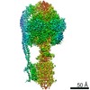
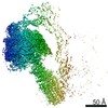
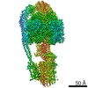
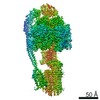
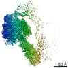
| Title | Cryo-EM structure of the mammalian DP-state ATP synthase | ||||||||||||
 Map data Map data | |||||||||||||
 Sample Sample |
| ||||||||||||
| Function / homology |  Function and homology information Function and homology informationFormation of ATP by chemiosmotic coupling / Cristae formation / : / mitochondrial proton-transporting ATP synthase complex binding / : / : / negative regulation of mitochondrial ATP synthesis coupled proton transport / Mitochondrial protein import / angiostatin binding / negative regulation of hydrolase activity ...Formation of ATP by chemiosmotic coupling / Cristae formation / : / mitochondrial proton-transporting ATP synthase complex binding / : / : / negative regulation of mitochondrial ATP synthesis coupled proton transport / Mitochondrial protein import / angiostatin binding / negative regulation of hydrolase activity / ATP biosynthetic process / mitochondrial depolarization / ATPase inhibitor activity / positive regulation of type 2 mitophagy / Mitochondrial translation termination / Mitochondrial protein degradation / positive regulation of mitochondrial outer membrane permeabilization involved in apoptotic signaling pathway / proton channel activity / heme biosynthetic process / negative regulation of endothelial cell proliferation / proton transmembrane transporter activity / proton motive force-driven ATP synthesis / proton-transporting two-sector ATPase complex, proton-transporting domain / proton motive force-driven mitochondrial ATP synthesis / H+-transporting two-sector ATPase / proton-transporting ATP synthase complex / proton-transporting ATP synthase activity, rotational mechanism / reactive oxygen species metabolic process / proton transmembrane transport / erythrocyte differentiation / ADP binding / mitochondrial membrane / ATPase binding / calmodulin binding / mitochondrial inner membrane / lipid binding / cell surface / protein-containing complex / mitochondrion / ATP binding / metal ion binding / identical protein binding / plasma membrane / cytoplasm Similarity search - Function | ||||||||||||
| Biological species |  | ||||||||||||
| Method | single particle reconstruction / cryo EM / Resolution: 3.34 Å | ||||||||||||
 Authors Authors | Gu JK / Zhang LX / Yi JB / Yang MJ | ||||||||||||
| Funding support |  China, 3 items China, 3 items
| ||||||||||||
 Citation Citation | Journal: Science / Year: 2019 Title: Cryo-EM structure of the mammalian ATP synthase tetramer bound with inhibitory protein IF1. Authors: Jinke Gu / Laixing Zhang / Shuai Zong / Runyu Guo / Tianya Liu / Jingbo Yi / Peiyi Wang / Wei Zhuo / Maojun Yang / Abstract: The mitochondrial adenosine triphosphate (ATP) synthase produces most of the ATP required by mammalian cells. We isolated porcine tetrameric ATP synthase and solved its structure at 6.2-angstrom ...The mitochondrial adenosine triphosphate (ATP) synthase produces most of the ATP required by mammalian cells. We isolated porcine tetrameric ATP synthase and solved its structure at 6.2-angstrom resolution using a single-particle cryo-electron microscopy method. Two classical V-shaped ATP synthase dimers lie antiparallel to each other to form an H-shaped ATP synthase tetramer, as viewed from the matrix. ATP synthase inhibitory factor subunit 1 (IF1) is a well-known in vivo inhibitor of mammalian ATP synthase at low pH. Two IF1 dimers link two ATP synthase dimers, which is consistent with the ATP synthase tetramer adopting an inhibited state. Within the tetramer, we refined structures of intact ATP synthase in two different rotational conformations at 3.34- and 3.45-Å resolution. | ||||||||||||
| History |
|
- Structure visualization
| Movie |
Movie viewer |
|---|---|
| Structure viewer | EM map: SurfViewMolmilJmol/JSmol |
| Supplemental images |

- Downloads & links
-EMDB archive
| Map data | emd_0677.map.gz | 5.5 MB | EMDB map data format | |
|---|---|---|---|---|
| Header (meta data) | emd-0677-v30.xmlemd-0677.xml | 8.3 KB 8.3 KB | Display Display | EMDB header |
| Images | 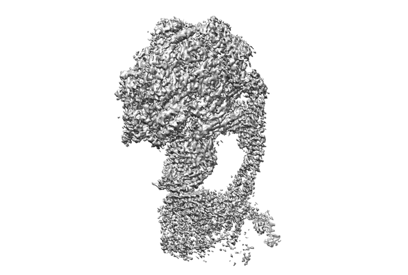 emd_0677.png emd_0677.png | 55.8 KB | ||
| Archive directory |  http://ftp.pdbj.org/pub/emdb/structures/EMD-0677ftp://ftp.pdbj.org/pub/emdb/structures/EMD-0677 http://ftp.pdbj.org/pub/emdb/structures/EMD-0677ftp://ftp.pdbj.org/pub/emdb/structures/EMD-0677 | HTTPS FTP |
-Related structure data
| Related structure data |  6j5iMC  0667C  0668C  0669C 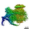 0670C 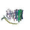 6j54C 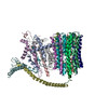 6j5aC 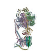 6j5jC 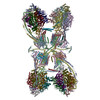 6j5kC M: atomic model generated by this map C: citing same article ( |
|---|---|
| Similar structure data | |
| EM raw data | EMPIAR-10283 (Title: Cryo-EM structure of the mammalian ATP synthase tetramer bound to inhibitory protein IF1 (Part1) Data size: 141.3 Data #1: Averaged micrographs of mammalian ATP synthase tetramer [micrographs - single frame]) |

-Links
| EMDB pages | EMDB (EBI/PDBe) / EMDataResource |
|---|---|
| Related items in Molecule of the Month |


-Map
| File | Download / File: emd_0677.map.gz / Format: CCP4 / Size: 52.7 MB / Type: IMAGE STORED AS FLOATING POINT NUMBER (4 BYTES) | ||||||||||||||||||||||||||||||||||||||||||||||||||||||||||||||||||||
|---|---|---|---|---|---|---|---|---|---|---|---|---|---|---|---|---|---|---|---|---|---|---|---|---|---|---|---|---|---|---|---|---|---|---|---|---|---|---|---|---|---|---|---|---|---|---|---|---|---|---|---|---|---|---|---|---|---|---|---|---|---|---|---|---|---|---|---|---|---|
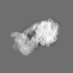
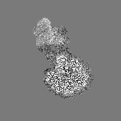
| Projections & slices | Image control
Images are generated by Spider. | ||||||||||||||||||||||||||||||||||||||||||||||||||||||||||||||||||||
| Voxel size | X=Y=Z: 1.38 Å | ||||||||||||||||||||||||||||||||||||||||||||||||||||||||||||||||||||
| Density |
| ||||||||||||||||||||||||||||||||||||||||||||||||||||||||||||||||||||
| Symmetry | Space group: 1 | ||||||||||||||||||||||||||||||||||||||||||||||||||||||||||||||||||||
| Details | EMDB XML:
CCP4 map header:
| ||||||||||||||||||||||||||||||||||||||||||||||||||||||||||||||||||||
 Z (Sec.)
 Y (Row.)
 X (Col.)


-Supplemental data
- Sample components
-Entire : Cryo-EM structure of the mammalian DP-state ATP synthase
| Entire | Name: Cryo-EM structure of the mammalian DP-state ATP synthase |
|---|---|
| Components |
|
-Supramolecule #1: Cryo-EM structure of the mammalian DP-state ATP synthase
| Supramolecule | Name: Cryo-EM structure of the mammalian DP-state ATP synthase type: complex / ID: 1 / Parent: 0 |
|---|---|
| Source (natural) | Organism: |
-Experimental details
-Structure determination
| Method | cryo EM |
|---|---|
 Processing Processing | single particle reconstruction |
| Aggregation state | particle |
-Sample preparation
| Buffer | pH: 7 |
|---|---|
| Vitrification | Cryogen name: ETHANE |
- Electron microscopy
| Microscope | FEI TITAN KRIOS |
|---|---|
| Image recording | Film or detector model: GATAN K2 SUMMIT (4k x 4k) / Average electron dose: 1.56 e/Å2 |
| Electron beam | Acceleration voltage: 300 kV / Electron source:  FIELD EMISSION GUN FIELD EMISSION GUN |
| Electron optics | Illumination mode: OTHER / Imaging mode: BRIGHT FIELD |
| Experimental equipment |  Model: Titan Krios / Image courtesy: FEI Company |
-Image processing
| Final reconstruction | Resolution.type: BY AUTHOR / Resolution: 3.34 Å / Resolution method: FSC 0.143 CUT-OFF / Number images used: 291128 |
|---|---|
| Initial angle assignment | Type: RANDOM ASSIGNMENT |
| Final angle assignment | Type: RANDOM ASSIGNMENT |
