 Movie
Movie Controller

[English] 日本語
 Yorodumi
- PDB-6itc: Structure of a substrate engaged SecA-SecY protein translocation ... -
+ Open data

- Basic information
| Entry | Database: PDB / ID: 6itc | |||||||||||||||||||||||||||||||||||||||
|---|---|---|---|---|---|---|---|---|---|---|---|---|---|---|---|---|---|---|---|---|---|---|---|---|---|---|---|---|---|---|---|---|---|---|---|---|---|---|---|---|
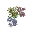
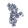
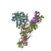
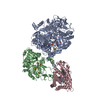
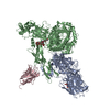
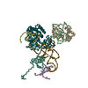
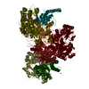
| Title | Structure of a substrate engaged SecA-SecY protein translocation machine | |||||||||||||||||||||||||||||||||||||||
 Components Components |
| |||||||||||||||||||||||||||||||||||||||
 Keywords Keywords | PROTEIN TRANSPORT / SecA / SecY / Translocation / Cryo-EM | |||||||||||||||||||||||||||||||||||||||
| Function / homology |  Function and homology information Function and homology informationouter membrane protein complex / cell envelope Sec protein transport complex / protein-exporting ATPase activity / protein-secreting ATPase / monoatomic ion transmembrane transporter activity / protein transport by the Sec complex / intracellular protein transmembrane transport / detection of virus / outer membrane / protein import ...outer membrane protein complex / cell envelope Sec protein transport complex / protein-exporting ATPase activity / protein-secreting ATPase / monoatomic ion transmembrane transporter activity / protein transport by the Sec complex / intracellular protein transmembrane transport / detection of virus / outer membrane / protein import / porin activity / pore complex / transmembrane protein transporter activity / protein secretion / protein targeting / monoatomic ion transport / bioluminescence / generation of precursor metabolites and energy / cell outer membrane / outer membrane-bounded periplasmic space / membrane raft / DNA damage response / symbiont entry into host cell / ATP binding / membrane / metal ion binding / identical protein binding / plasma membrane / cytoplasm Similarity search - Function | |||||||||||||||||||||||||||||||||||||||
| Biological species |  Geobacillus thermodenitrificans (bacteria) Geobacillus thermodenitrificans (bacteria)   Aequorea victoria (jellyfish) Aequorea victoria (jellyfish) | |||||||||||||||||||||||||||||||||||||||
| Method | ELECTRON MICROSCOPY / single particle reconstruction / cryo EM / Resolution: 3.45 Å | |||||||||||||||||||||||||||||||||||||||
 Authors Authors | Ma, C.Y. / Wu, X.F. / Sun, D.J. / Park, E.Y. / Rapoport, T.A. / Gao, N. / Long, L. | |||||||||||||||||||||||||||||||||||||||
| Funding support |  China, 1items China, 1items
| |||||||||||||||||||||||||||||||||||||||
 Citation Citation | Journal: Nat Commun / Year: 2019 Title: Structure of the substrate-engaged SecA-SecY protein translocation machine. Authors: Chengying Ma / Xiaofei Wu / Dongjie Sun / Eunyong Park / Marco A Catipovic / Tom A Rapoport / Ning Gao / Long Li /  Abstract: The Sec61/SecY channel allows the translocation of many proteins across the eukaryotic endoplasmic reticulum membrane or the prokaryotic plasma membrane. In bacteria, most secretory proteins are ...The Sec61/SecY channel allows the translocation of many proteins across the eukaryotic endoplasmic reticulum membrane or the prokaryotic plasma membrane. In bacteria, most secretory proteins are transported post-translationally through the SecY channel by the SecA ATPase. How a polypeptide is moved through the SecA-SecY complex is poorly understood, as structural information is lacking. Here, we report an electron cryo-microscopy (cryo-EM) structure of a translocating SecA-SecY complex in a lipid environment. The translocating polypeptide chain can be traced through both SecA and SecY. In the captured transition state of ATP hydrolysis, SecA's two-helix finger is close to the polypeptide, while SecA's clamp interacts with the polypeptide in a sequence-independent manner by inducing a short β-strand. Taking into account previous biochemical and biophysical data, our structure is consistent with a model in which the two-helix finger and clamp cooperate during the ATPase cycle to move a polypeptide through the channel. | |||||||||||||||||||||||||||||||||||||||
| History |
|
- Structure visualization
| Movie |
Movie viewer |
|---|---|
| Structure viewer | Molecule: MolmilJmol/JSmol |
- Downloads & links
-Download
| PDBx/mmCIF format | 6itc.cif.gz | 322.5 KB | Display | PDBx/mmCIF format |
|---|---|---|---|---|
| PDB format | pdb6itc.ent.gz | 253.7 KB | Display | PDB format |
| PDBx/mmJSON format | 6itc.json.gz | Tree view | PDBx/mmJSON format | |
| Others |  Other downloads Other downloads |
-Validation report
| Arichive directory | https://data.pdbj.org/pub/pdb/validation_reports/it/6itcftp://data.pdbj.org/pub/pdb/validation_reports/it/6itc | HTTPS FTP |
|---|
-Related structure data
| Related structure data | 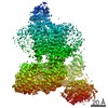 9731MC M: map data used to model this data C: citing same article ( |
|---|---|
| Similar structure data |

-Links
 PDBj
PDBj


- Assembly
| Deposited unit | 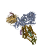
|
|---|---|
| 1 |
|
-Components
-Protein translocase subunit ... , 3 types, 3 molecules AYE
| #1: Protein | Mass: 88916.180 Da / Num. of mol.: 1 Source method: isolated from a genetically manipulated source Source: (gene. exp.) Strain: 168 / Gene: secA, div+, BSU35300 / Production host: |
|---|---|
| #2: Protein | Mass: 46768.301 Da / Num. of mol.: 1 / Mutation: G60C,Q202T,L210G,F211G,R213N Source method: isolated from a genetically manipulated source Source: (gene. exp.) Geobacillus thermodenitrificans (strain NG80-2) (bacteria)Strain: NG80-2 / Gene: secY, GTNG_0125 / Production host: |
| #3: Protein | Mass: 8249.600 Da / Num. of mol.: 1 Source method: isolated from a genetically manipulated source Source: (gene. exp.) Geobacillus thermodenitrificans (strain NG80-2) (bacteria)Strain: NG80-2 / Gene: secE, GTNG_0091 / Production host: |
-Protein , 2 types, 2 molecules BG
| #5: Protein | Mass: 6024.562 Da / Num. of mol.: 1 Source method: isolated from a genetically manipulated source Source: (gene. exp.) |
|---|---|
| #6: Protein | Mass: 26813.113 Da / Num. of mol.: 1 / Mutation: Q80R,F99S,M153T,V163A Source method: isolated from a genetically manipulated source Source: (gene. exp.) Aequorea victoria (jellyfish) / Gene: GFP / Production host: |
-Antibody , 2 types, 2 molecules VC
| #4: Antibody | Mass: 12919.544 Da / Num. of mol.: 1 Source method: isolated from a genetically manipulated source Source: (gene. exp.) |
|---|---|
| #7: Antibody | Mass: 12368.727 Da / Num. of mol.: 1 Source method: isolated from a genetically manipulated source Source: (gene. exp.) |
-Non-polymers , 4 types, 5 molecules 


| #8: Chemical | ChemComp-MG /  Mass: 24.305 Da / Num. of mol.: 1 / Source method: obtained synthetically / Formula: Mg Mass: 24.305 Da / Num. of mol.: 1 / Source method: obtained synthetically / Formula: Mg |
|---|---|
| #9: Chemical | ChemComp-BEF /  Mass: 66.007 Da / Num. of mol.: 1 / Source method: obtained synthetically / Formula: BeF3 Mass: 66.007 Da / Num. of mol.: 1 / Source method: obtained synthetically / Formula: BeF3 |
| #10: Chemical | ChemComp-ADP /  Mass: 427.201 Da / Num. of mol.: 1 / Source method: obtained synthetically / Formula: C10H15N5O10P2 / Comment: ADP, energy-carrying molecule*YM Mass: 427.201 Da / Num. of mol.: 1 / Source method: obtained synthetically / Formula: C10H15N5O10P2 / Comment: ADP, energy-carrying molecule*YM |
| #11: Chemical |  Mass: 749.007 Da / Num. of mol.: 2 / Source method: obtained synthetically / Formula: C40H77O10P / Comment: phospholipid*YM Mass: 749.007 Da / Num. of mol.: 2 / Source method: obtained synthetically / Formula: C40H77O10P / Comment: phospholipid*YM |
-Details
| Has protein modification | Y |
|---|
-Experimental details
-Experiment
| Experiment | Method: ELECTRON MICROSCOPY |
|---|---|
| EM experiment | Aggregation state: PARTICLE / 3D reconstruction method: single particle reconstruction |
- Sample preparation
| Component | Name: SecA-SecY complex / Type: COMPLEX / Entity ID: #1-#7 / Source: RECOMBINANT |
|---|---|
| Source (natural) | Organism: |
| Source (recombinant) | Organism: |
| Buffer solution | pH: 7.5 |
| Specimen | Embedding applied: NO / Shadowing applied: NO / Staining applied: NO / Vitrification applied: YES |
| Vitrification | Cryogen name: ETHANE |
- Electron microscopy imaging
| Experimental equipment |  Model: Titan Krios / Image courtesy: FEI Company |
|---|---|
| Microscopy | Model: FEI TITAN KRIOS |
| Electron gun | Electron source:  FIELD EMISSION GUN / Accelerating voltage: 300 kV / Illumination mode: FLOOD BEAM FIELD EMISSION GUN / Accelerating voltage: 300 kV / Illumination mode: FLOOD BEAM |
| Electron lens | Mode: BRIGHT FIELD |
| Image recording | Electron dose: 5 e/Å2 / Film or detector model: GATAN K2 QUANTUM (4k x 4k) |
- Processing
| Software | Name: PHENIX / Version: 1.13_2998: / Classification: refinement |
|---|---|
| EM software | Name: PHENIX / Category: model refinement |
| CTF correction | Type: PHASE FLIPPING AND AMPLITUDE CORRECTION |
| Symmetry | Point symmetry: C1 (asymmetric) |
| 3D reconstruction | Resolution: 3.45 Å / Resolution method: FSC 0.143 CUT-OFF / Num. of particles: 130153 / Symmetry type: POINT |
| Atomic model building | Protocol: FLEXIBLE FIT |
