Movie
Movie Controller


+ Open data

- Basic information
| Entry | Database: PDB / ID: 6hwy | ||||||||||||||||||||||||||||||
|---|---|---|---|---|---|---|---|---|---|---|---|---|---|---|---|---|---|---|---|---|---|---|---|---|---|---|---|---|---|---|---|
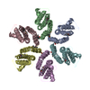
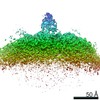
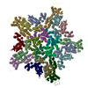
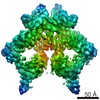
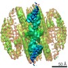
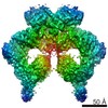
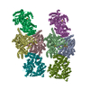
| Title | Mature MLV capsid pentamer structure in intact virus particles | ||||||||||||||||||||||||||||||
 Components Components | Putative gag polyprotein | ||||||||||||||||||||||||||||||
 Keywords Keywords | VIRAL PROTEIN / MLV / capsid / pentamer | ||||||||||||||||||||||||||||||
| Function / homology |  Function and homology information Function and homology informationvirion assembly / viral budding via host ESCRT complex / host multivesicular body / viral nucleocapsid / structural constituent of virion / nucleic acid binding / host cell plasma membrane / RNA binding / zinc ion binding Similarity search - Function | ||||||||||||||||||||||||||||||
| Biological species |  Murine leukemia virus Murine leukemia virus | ||||||||||||||||||||||||||||||
| Method | ELECTRON MICROSCOPY / subtomogram averaging / cryo EM / Resolution: 8.6 Å | ||||||||||||||||||||||||||||||
 Authors Authors | Qu, K. / Glass, B. / Dolezal, M. / Schur, F.K.M. / Rein, A. / Rumlova, M. / Ruml, T. / Kraeusslich, H.G. / Briggs, J.A.G. | ||||||||||||||||||||||||||||||
| Funding support |  Germany, Germany,  United Kingdom, United Kingdom,  Czech Republic, 9items Czech Republic, 9items
| ||||||||||||||||||||||||||||||
 Citation Citation | Journal: Proc Natl Acad Sci U S A / Year: 2018 Title: Structure and architecture of immature and mature murine leukemia virus capsids. Authors: Kun Qu / Bärbel Glass / Michal Doležal / Florian K M Schur / Brice Murciano / Alan Rein / Michaela Rumlová / Tomáš Ruml / Hans-Georg Kräusslich / John A G Briggs /   Abstract: Retroviruses assemble and bud from infected cells in an immature form and require proteolytic maturation for infectivity. The CA (capsid) domains of the Gag polyproteins assemble a protein lattice as ...Retroviruses assemble and bud from infected cells in an immature form and require proteolytic maturation for infectivity. The CA (capsid) domains of the Gag polyproteins assemble a protein lattice as a truncated sphere in the immature virion. Proteolytic cleavage of Gag induces dramatic structural rearrangements; a subset of cleaved CA subsequently assembles into the mature core, whose architecture varies among retroviruses. Murine leukemia virus (MLV) is the prototypical γ-retrovirus and serves as the basis of retroviral vectors, but the structure of the MLV CA layer is unknown. Here we have combined X-ray crystallography with cryoelectron tomography to determine the structures of immature and mature MLV CA layers within authentic viral particles. This reveals the structural changes associated with maturation, and, by comparison with HIV-1, uncovers conserved and variable features. In contrast to HIV-1, most MLV CA is used for assembly of the mature core, which adopts variable, multilayered morphologies and does not form a closed structure. Unlike in HIV-1, there is similarity between protein-protein interfaces in the immature MLV CA layer and those in the mature CA layer, and structural maturation of MLV could be achieved through domain rotations that largely maintain hexameric interactions. Nevertheless, the dramatic architectural change on maturation indicates that extensive disassembly and reassembly are required for mature core growth. The core morphology suggests that wrapping of the genome in CA sheets may be sufficient to protect the MLV ribonucleoprotein during cell entry. | ||||||||||||||||||||||||||||||
| History |
|
- Structure visualization
| Movie |
Movie viewer |
|---|---|
| Structure viewer | Molecule: MolmilJmol/JSmol |
- Downloads & links
-Download
| PDBx/mmCIF format | 6hwy.cif.gz | 146.2 KB | Display | PDBx/mmCIF format |
|---|---|---|---|---|
| PDB format | pdb6hwy.ent.gz | 98.5 KB | Display | PDB format |
| PDBx/mmJSON format | 6hwy.json.gz | Tree view | PDBx/mmJSON format | |
| Others |  Other downloads Other downloads |
-Validation report
| Arichive directory | https://data.pdbj.org/pub/pdb/validation_reports/hw/6hwyftp://data.pdbj.org/pub/pdb/validation_reports/hw/6hwy | HTTPS FTP |
|---|
-Related structure data
| Related structure data | 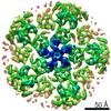 0293MC 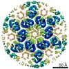 0290C 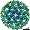 0291C 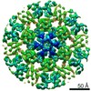 0292C  4419C  4421C  4422C 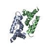 6gzaC 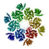 6hwiC 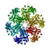 6hwwC 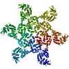 6hwxC M: map data used to model this data C: citing same article ( |
|---|---|
| Similar structure data |
-Links
 PDBj
PDBj- Assembly
| Deposited unit | 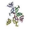
|
|---|---|
| 1 | x 5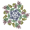
|
-Components
| #1: Protein | Mass: 27165.305 Da / Num. of mol.: 4 Source method: isolated from a genetically manipulated source Source: (gene. exp.) Murine leukemia virus / Gene: gag / Cell line (production host): HEK293T / Production host:  Homo sapiens (human) / References: UniProt: A0A240FAQ8, UniProt: P03336*PLUS Homo sapiens (human) / References: UniProt: A0A240FAQ8, UniProt: P03336*PLUS |
|---|
-Experimental details
-Experiment
| Experiment | Method: ELECTRON MICROSCOPY |
|---|---|
| EM experiment | Aggregation state: PARTICLE / 3D reconstruction method: subtomogram averaging |
- Sample preparation
| Component | Name: Murine leukemia virus / Type: VIRUS / Entity ID: all / Source: RECOMBINANT |
|---|---|
| Source (natural) | Organism: Murine leukemia virus |
| Source (recombinant) | Organism: Homo sapiens (human) / Cell: HEK 293T / Plasmid: M2204 |
| Details of virus | Empty: NO / Enveloped: YES / Isolate: STRAIN / Type: VIRION |
| Buffer solution | pH: 6 |
| Specimen | Embedding applied: NO / Shadowing applied: NO / Staining applied: NO / Vitrification applied: YES Details: Purified virus solution was inactivated and diluted 1:1 with PBS containing 10 nm colloidal gold. |
| Specimen support | Grid material: COPPER / Grid mesh size: 300 divisions/in. / Grid type: C-flat-2/1 |
| Vitrification | Instrument: FEI VITROBOT MARK II / Cryogen name: ETHANE / Humidity: 95 % / Chamber temperature: 288 K |
- Electron microscopy imaging
| Experimental equipment |  Model: Titan Krios / Image courtesy: FEI Company |
|---|---|
| Microscopy | Model: FEI TITAN KRIOS |
| Electron gun | Electron source:  FIELD EMISSION GUN / Accelerating voltage: 300 kV / Illumination mode: FLOOD BEAM FIELD EMISSION GUN / Accelerating voltage: 300 kV / Illumination mode: FLOOD BEAM |
| Electron lens | Mode: BRIGHT FIELD / Nominal magnification: 105000 X / Nominal defocus max: 7500 nm / Nominal defocus min: 2000 nm / Cs: 2.7 mm / C2 aperture diameter: 50 µm / Alignment procedure: ZEMLIN TABLEAU |
| Specimen holder | Cryogen: NITROGEN / Specimen holder model: FEI TITAN KRIOS AUTOGRID HOLDER |
| Image recording | Average exposure time: 0.6 sec. / Electron dose: 1.8 e/Å2 / Detector mode: SUPER-RESOLUTION / Film or detector model: GATAN K2 QUANTUM (4k x 4k) / Num. of grids imaged: 2 Details: Dose fluctuation was caused by the ring collapse of FEG during data collection. |
| EM imaging optics | Energyfilter name: GIF Quantum LS / Energyfilter slit width: 20 eV |
| Image scans | Sampling size: 5 µm / Width: 3710 / Height: 3838 / Movie frames/image: 6 / Used frames/image: 1-6 |
- Processing
| EM software |
| ||||||||||||||||||||||||||||||||||||||||||||||||
|---|---|---|---|---|---|---|---|---|---|---|---|---|---|---|---|---|---|---|---|---|---|---|---|---|---|---|---|---|---|---|---|---|---|---|---|---|---|---|---|---|---|---|---|---|---|---|---|---|---|
| CTF correction | Type: PHASE FLIPPING ONLY | ||||||||||||||||||||||||||||||||||||||||||||||||
| Symmetry | Point symmetry: C5 (5 fold cyclic) | ||||||||||||||||||||||||||||||||||||||||||||||||
| 3D reconstruction | Resolution: 8.6 Å / Resolution method: FSC 0.143 CUT-OFF / Num. of particles: 1299 / Algorithm: BACK PROJECTION / Symmetry type: POINT | ||||||||||||||||||||||||||||||||||||||||||||||||
| EM volume selection | Num. of tomograms: 65 / Num. of volumes extracted: 1299 | ||||||||||||||||||||||||||||||||||||||||||||||||
| Atomic model building | Protocol: RIGID BODY FIT / Space: REAL | ||||||||||||||||||||||||||||||||||||||||||||||||
| Atomic model building | 3D fitting-ID: 1 / Pdb chain-ID: A / Source name: PDB / Type: experimental model
|