 Movie
Movie Controller

[English] 日本語
 Yorodumi
- PDB-6dzt: Cryo-EM structure of nucleosome in complex with a single chain an... -
+ Open data

- Basic information
| Entry | Database: PDB / ID: 6dzt | ||||||
|---|---|---|---|---|---|---|---|
| Title | Cryo-EM structure of nucleosome in complex with a single chain antibody fragment | ||||||
 Components Components |
| ||||||
 Keywords Keywords | NUCLEAR PROTEIN / Nucleosome / Single chain antibody / charge-charge interaction / acidic patch. | ||||||
| Function / homology |  Function and homology information Function and homology informationHDMs demethylate histones / PKMTs methylate histone lysines / Interleukin-7 signaling / Chromatin modifying enzymes / : / SUMOylation of chromatin organization proteins / Metalloprotease DUBs / E3 ubiquitin ligases ubiquitinate target proteins / Factors involved in megakaryocyte development and platelet production / RCAF complex ...HDMs demethylate histones / PKMTs methylate histone lysines / Interleukin-7 signaling / Chromatin modifying enzymes / : / SUMOylation of chromatin organization proteins / Metalloprotease DUBs / E3 ubiquitin ligases ubiquitinate target proteins / Factors involved in megakaryocyte development and platelet production / RCAF complex / RMTs methylate histone arginines / Recruitment and ATM-mediated phosphorylation of repair and signaling proteins at DNA double strand breaks / SIRT1 negatively regulates rRNA expression / NoRC negatively regulates rRNA expression / Activated PKN1 stimulates transcription of AR (androgen receptor) regulated genes KLK2 and KLK3 / polytene chromosome band / PRC2 methylates histones and DNA / HDACs deacetylate histones / Ub-specific processing proteases / Negative Regulation of CDH1 Gene Transcription / Formation of the beta-catenin:TCF transactivating complex / MLL4 and MLL3 complexes regulate expression of PPARG target genes in adipogenesis and hepatic steatosis / RNA Polymerase I Promoter Escape / Regulation of endogenous retroelements by KRAB-ZFP proteins / larval somatic muscle development / RUNX1 regulates genes involved in megakaryocyte differentiation and platelet function / Senescence-Associated Secretory Phenotype (SASP) / Transcriptional regulation by small RNAs / Estrogen-dependent gene expression / HATs acetylate histones / Assembly of the ORC complex at the origin of replication / Oxidative Stress Induced Senescence / UCH proteinases / polytene chromosome / nucleosomal DNA binding / nuclear chromosome / structural constituent of chromatin / nucleosome / nucleosome assembly / heterochromatin formation / chromosome / chromatin organization / protein heterodimerization activity / chromatin / protein-containing complex binding / DNA binding / nucleus Similarity search - Function | ||||||
| Biological species |    | ||||||
| Method | ELECTRON MICROSCOPY / single particle reconstruction / cryo EM / Resolution: 2.99 Å | ||||||
 Authors Authors | Yadav, K.N.S. / Zhou, B.-R. | ||||||
| Funding support |  United States, 1items United States, 1items
| ||||||
 Citation Citation | Journal: Nat Commun / Year: 2019 Title: Atomic resolution cryo-EM structure of a native-like CENP-A nucleosome aided by an antibody fragment. Authors: Bing-Rui Zhou / K N Sathish Yadav / Mario Borgnia / Jingjun Hong / Baohua Cao / Ada L Olins / Donald E Olins / Yawen Bai / Ping Zhang / Abstract: Genomic DNA in eukaryotes is organized into chromatin through association with core histones to form nucleosomes, each distinguished by their DNA sequences and histone variants. Here, we used a ...Genomic DNA in eukaryotes is organized into chromatin through association with core histones to form nucleosomes, each distinguished by their DNA sequences and histone variants. Here, we used a single-chain antibody fragment (scFv) derived from the anti-nucleosome antibody mAb PL2-6 to stabilize human CENP-A nucleosome containing a native α-satellite DNA and solved its structure by the cryo-electron microscopy (cryo-EM) to 2.6 Å resolution. In comparison, the corresponding cryo-EM structure of the free CENP-A nucleosome could only reach 3.4 Å resolution. We find that scFv binds to a conserved acidic patch on the histone H2A-H2B dimer without perturbing the nucleosome structure. Our results provide an atomic resolution cryo-EM structure of a nucleosome and insight into the structure and function of the CENP-A nucleosome. The scFv approach is applicable to the structural determination of other native-like nucleosomes with distinct DNA sequences. | ||||||
| History |
|
- Structure visualization
| Movie |
Movie viewer |
|---|---|
| Structure viewer | Molecule: MolmilJmol/JSmol |
- Downloads & links
-Download
| PDBx/mmCIF format | 6dzt.cif.gz | 364.7 KB | Display | PDBx/mmCIF format |
|---|---|---|---|---|
| PDB format | pdb6dzt.ent.gz | 278.5 KB | Display | PDB format |
| PDBx/mmJSON format | 6dzt.json.gz | Tree view | PDBx/mmJSON format | |
| Others |  Other downloads Other downloads |
-Validation report
| Arichive directory | https://data.pdbj.org/pub/pdb/validation_reports/dz/6dztftp://data.pdbj.org/pub/pdb/validation_reports/dz/6dzt | HTTPS FTP |
|---|
-Related structure data
| Related structure data | 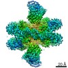 8938MC 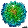 0586C 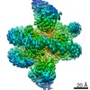 8945C 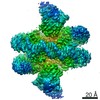 8949C  6e0cC  6e0pC  6o1dC M: map data used to model this data C: citing same article ( |
|---|---|
| Similar structure data |

-Links
 PDBj
PDBj


- Assembly
| Deposited unit | 
|
|---|---|
| 1 |
|
-Components
-Protein , 4 types, 8 molecules AEBFCGDH
| #1: Protein | Mass: 15421.101 Da / Num. of mol.: 2 Source method: isolated from a genetically manipulated source Source: (gene. exp.) Gene: His3, His3:CG31613, CG31613, His3:CG33803, CG33803, His3:CG33806, CG33806, His3:CG33809, CG33809, His3:CG33812, CG33812, His3:CG33815, CG33815, His3:CG33818, CG33818, His3:CG33821, CG33821, ...Gene: His3, His3:CG31613, CG31613, His3:CG33803, CG33803, His3:CG33806, CG33806, His3:CG33809, CG33809, His3:CG33812, CG33812, His3:CG33815, CG33815, His3:CG33818, CG33818, His3:CG33821, CG33821, His3:CG33824, CG33824, His3:CG33827, CG33827, His3:CG33830, CG33830, His3:CG33833, CG33833, His3:CG33836, CG33836, His3:CG33839, CG33839, His3:CG33842, CG33842, His3:CG33845, CG33845, His3:CG33848, CG33848, His3:CG33851, CG33851, His3:CG33854, CG33854, His3:CG33857, CG33857, His3:CG33860, CG33860, His3:CG33863, CG33863, His3:CG33866, CG33866 Production host: #2: Protein | Mass: 11521.611 Da / Num. of mol.: 2 Source method: isolated from a genetically manipulated source Source: (gene. exp.) Gene: His4, H4, His4r, H4r, CG3379, His4:CG31611, CG31611, His4:CG33869, CG33869, His4:CG33871, CG33871, His4:CG33873, CG33873, His4:CG33875, CG33875, His4:CG33877, CG33877, His4:CG33879, CG33879, ...Gene: His4, H4, His4r, H4r, CG3379, His4:CG31611, CG31611, His4:CG33869, CG33869, His4:CG33871, CG33871, His4:CG33873, CG33873, His4:CG33875, CG33875, His4:CG33877, CG33877, His4:CG33879, CG33879, His4:CG33881, CG33881, His4:CG33883, CG33883, His4:CG33885, CG33885, His4:CG33887, CG33887, His4:CG33889, CG33889, His4:CG33891, CG33891, His4:CG33893, CG33893, His4:CG33895, CG33895, His4:CG33897, CG33897, His4:CG33899, CG33899, His4:CG33901, CG33901, His4:CG33903, CG33903, His4:CG33905, CG33905, His4:CG33907, CG33907, His4:CG33909, CG33909 Production host: #3: Protein | Mass: 13388.727 Da / Num. of mol.: 2 Source method: isolated from a genetically manipulated source Source: (gene. exp.) Gene: His2A, H2a, His2A:CG31618, CG31618, His2A:CG33808, CG33808, His2A:CG33814, CG33814, His2A:CG33817, CG33817, His2A:CG33820, CG33820, His2A:CG33823, CG33823, His2A:CG33826, CG33826, His2A: ...Gene: His2A, H2a, His2A:CG31618, CG31618, His2A:CG33808, CG33808, His2A:CG33814, CG33814, His2A:CG33817, CG33817, His2A:CG33820, CG33820, His2A:CG33823, CG33823, His2A:CG33826, CG33826, His2A:CG33829, CG33829, His2A:CG33832, CG33832, His2A:CG33835, CG33835, His2A:CG33838, CG33838, His2A:CG33841, CG33841, His2A:CG33844, CG33844, His2A:CG33847, CG33847, His2A:CG33850, CG33850, His2A:CG33862, CG33862, His2A:CG33865, CG33865 Production host: #4: Protein | Mass: 13840.224 Da / Num. of mol.: 2 Source method: isolated from a genetically manipulated source Source: (gene. exp.) Gene: His2B, His2B:CG17949, CG17949, His2B:CG33868, CG33868, His2B:CG33870, CG33870, His2B:CG33872, CG33872, His2B:CG33874, CG33874, His2B:CG33876, CG33876, His2B:CG33878, CG33878, His2B:CG33880, ...Gene: His2B, His2B:CG17949, CG17949, His2B:CG33868, CG33868, His2B:CG33870, CG33870, His2B:CG33872, CG33872, His2B:CG33874, CG33874, His2B:CG33876, CG33876, His2B:CG33878, CG33878, His2B:CG33880, CG33880, His2B:CG33882, CG33882, His2B:CG33884, CG33884, His2B:CG33886, CG33886, His2B:CG33888, CG33888, His2B:CG33890, CG33890, His2B:CG33892, CG33892, His2B:CG33894, CG33894, His2B:CG33896, CG33896, His2B:CG33898, CG33898, His2B:CG33900, CG33900, His2B:CG33902, CG33902, His2B:CG33904, CG33904, His2B:CG33906, CG33906, His2B:CG33908, CG33908, His2B:CG33910, CG33910 Production host: |
|---|
-DNA chain , 2 types, 2 molecules IJ
| #5: DNA chain | Mass: 45610.043 Da / Num. of mol.: 1 / Source method: obtained synthetically / Source: (synth.) |
|---|---|
| #6: DNA chain | Mass: 45138.770 Da / Num. of mol.: 1 / Source method: obtained synthetically / Source: (synth.) |
-Antibody , 1 types, 2 molecules MN
| #7: Antibody | Mass: 29030.146 Da / Num. of mol.: 2 Source method: isolated from a genetically manipulated source Source: (gene. exp.) |
|---|
-Details
| Has protein modification | Y |
|---|
-Experimental details
-Experiment
| Experiment | Method: ELECTRON MICROSCOPY |
|---|---|
| EM experiment | Aggregation state: PARTICLE / 3D reconstruction method: single particle reconstruction |
- Sample preparation
| Component | Name: Nucleosome-Antibody complex / Type: COMPLEX / Entity ID: all / Source: RECOMBINANT |
|---|---|
| Source (natural) | Organism: |
| Source (recombinant) | Organism: |
| Buffer solution | pH: 7.4 |
| Specimen | Embedding applied: NO / Shadowing applied: NO / Staining applied: NO / Vitrification applied: YES |
| Vitrification | Cryogen name: ETHANE |
- Electron microscopy imaging
| Experimental equipment |  Model: Titan Krios / Image courtesy: FEI Company |
|---|---|
| Microscopy | Model: FEI TITAN KRIOS |
| Electron gun | Electron source:  FIELD EMISSION GUN / Accelerating voltage: 300 kV / Illumination mode: OTHER FIELD EMISSION GUN / Accelerating voltage: 300 kV / Illumination mode: OTHER |
| Electron lens | Mode: OTHER |
| Image recording | Electron dose: 40 e/Å2 / Film or detector model: GATAN K2 SUMMIT (4k x 4k) |
- Processing
| Software |
| ||||||||||||||||||||||||
|---|---|---|---|---|---|---|---|---|---|---|---|---|---|---|---|---|---|---|---|---|---|---|---|---|---|
| EM software | Name: RELION / Version: 3.0 beta / Category: 3D reconstruction | ||||||||||||||||||||||||
| CTF correction | Type: PHASE FLIPPING ONLY | ||||||||||||||||||||||||
| Symmetry | Point symmetry: C1 (asymmetric) | ||||||||||||||||||||||||
| 3D reconstruction | Resolution: 2.99 Å / Resolution method: FSC 0.143 CUT-OFF / Num. of particles: 238790 / Num. of class averages: 2 / Symmetry type: POINT | ||||||||||||||||||||||||
| Refinement | Stereochemistry target values: GeoStd + Monomer Library | ||||||||||||||||||||||||
| Refine LS restraints |
|
