National Institutes of Health/National Institute of Diabetes and Digestive and Kidney Disease (NIH/NIDDK)
R01 DK110575-01
米国
引用
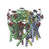
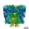
ジャーナル: Nat Commun / 年: 2018 タイトル: Hydrophobic pore gates regulate ion permeation in polycystic kidney disease 2 and 2L1 channels. 著者: Wang Zheng / Xiaoyong Yang / Ruikun Hu / Ruiqi Cai / Laura Hofmann / Zhifei Wang / Qiaolin Hu / Xiong Liu / David Bulkley / Yong Yu / Jingfeng Tang / Veit Flockerzi / Ying Cao / Erhu Cao / Xing-Zhen Chen / 要旨: PKD2 and PKD1 genes are mutated in human autosomal dominant polycystic kidney disease. PKD2 can form either a homomeric cation channel or a heteromeric complex with the PKD1 receptor, presumed to ...PKD2 and PKD1 genes are mutated in human autosomal dominant polycystic kidney disease. PKD2 can form either a homomeric cation channel or a heteromeric complex with the PKD1 receptor, presumed to respond to ligand(s) and/or mechanical stimuli. Here, we identify a two-residue hydrophobic gate in PKD2L1, and a single-residue hydrophobic gate in PKD2. We find that a PKD2 gain-of-function gate mutant effectively rescues PKD2 knockdown-induced phenotypes in embryonic zebrafish. The structure of a PKD2 activating mutant F604P by cryo-electron microscopy reveals a π- to α-helix transition within the pore-lining helix S6 that leads to repositioning of the gate residue and channel activation. Overall the results identify hydrophobic gates and a gating mechanism of PKD2 and PKD2L1.
履歴
登録
2018年4月12日
登録サイト: RCSB / 処理サイト: RCSB
改定 1.0
2018年6月27日
Provider: repository / タイプ: Initial release
改定 1.0
2018年6月27日
Data content type: EM metadata / Data content type: EM metadata / Provider: repository / タイプ: Initial release
改定 1.0
2018年6月27日
Data content type: Half map / Part number: 1 / Data content type: Half map / Provider: repository / タイプ: Initial release
改定 1.0
2018年6月27日
Data content type: Half map / Part number: 2 / Data content type: Half map / Provider: repository / タイプ: Initial release
改定 1.0
2018年6月27日
Data content type: Image / Data content type: Image / Provider: repository / タイプ: Initial release
改定 1.0
2018年6月27日
Data content type: Primary map / Data content type: Primary map / Provider: repository / タイプ: Initial release
改定 1.0
2018年6月27日
Data content type: Half map / Part number: 1 / Data content type: Half map / Provider: repository / タイプ: Initial release
改定 1.0
2018年6月27日
Data content type: Half map / Part number: 2 / Data content type: Half map / Provider: repository / タイプ: Initial release
改定 1.0
2018年6月27日
Data content type: Image / Data content type: Image / Provider: repository / タイプ: Initial release
改定 1.0
2018年6月27日
Data content type: Primary map / Data content type: Primary map / Provider: repository / タイプ: Initial release
Data content type: EM metadata / Data content type: EM metadata / EM metadata / Group: Data processing / Experimental summary / Data content type: EM metadata / EM metadata / カテゴリ: em_admin / em_software / Data content type: EM metadata / EM metadata / Item: _em_admin.last_update / _em_software.name
 ムービー
ムービー コントローラー


 データを開く
 基本情報
基本情報 要素
要素 キーワード
キーワード 機能・相同性情報
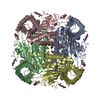
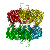
 Homo sapiens (ヒト)
 データ登録者
 米国, 1件
  引用
引用


 構造の表示
構造の表示 ダウンロードとリンク
 その他のダウンロード


 PDBj
PDBj
 集合体
集合体

 タイプ: D-saccharide, beta linking / 分子量: 221.208 Da / 分子数: 12 / 由来タイプ: 組換発現 / 式: C8H15NO6
 試料調製
試料調製 電子顕微鏡撮影
 FIELD EMISSION GUN / 加速電圧: 300 kV / 照射モード: FLOOD BEAM
 解析
解析