 Movie
Movie Controller

[English] 日本語
 Yorodumi
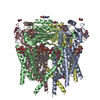
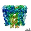
- PDB-5mkf: cryoEM Structure of Polycystin-2 in complex with calcium and lipids -
+ Open data

- Basic information
| Entry | Database: PDB / ID: 5mkf | |||||||||
|---|---|---|---|---|---|---|---|---|---|---|
| Title | cryoEM Structure of Polycystin-2 in complex with calcium and lipids | |||||||||
 Components Components | Polycystin-2 | |||||||||
 Keywords Keywords | TRANSPORT PROTEIN / Ca2+ signaling / cryoEM / membrane protein structure / Polycystin-2 / TRP channel | |||||||||
| Function / homology |  Function and homology information Function and homology informationdetection of nodal flow / metanephric smooth muscle tissue development / metanephric cortex development / metanephric cortical collecting duct development / metanephric distal tubule development / polycystin complex / mesonephric tubule development / mesonephric duct development / metanephric part of ureteric bud development / renal tubule morphogenesis ...detection of nodal flow / metanephric smooth muscle tissue development / metanephric cortex development / metanephric cortical collecting duct development / metanephric distal tubule development / polycystin complex / mesonephric tubule development / mesonephric duct development / metanephric part of ureteric bud development / renal tubule morphogenesis / determination of liver left/right asymmetry / metanephric ascending thin limb development / metanephric mesenchyme development / metanephric S-shaped body morphogenesis / basal cortex / placenta blood vessel development / renal artery morphogenesis / HLH domain binding / VxPx cargo-targeting to cilium / cilium organization / migrasome / cellular response to fluid shear stress / muscle alpha-actinin binding / regulation of calcium ion import / calcium-induced calcium release activity / detection of mechanical stimulus / determination of left/right symmetry / voltage-gated monoatomic ion channel activity / cellular response to hydrostatic pressure / aorta development / cation channel complex / branching involved in ureteric bud morphogenesis / neural tube development / non-motile cilium / outward rectifier potassium channel activity / motile cilium / actinin binding / cellular response to osmotic stress / voltage-gated sodium channel activity / negative regulation of G1/S transition of mitotic cell cycle / ciliary membrane / voltage-gated monoatomic cation channel activity / heart looping / positive regulation of phospholipase C-activating G protein-coupled receptor signaling pathway / protein heterotetramerization / spinal cord development / embryonic placenta development / cytoplasmic side of endoplasmic reticulum membrane / centrosome duplication / voltage-gated potassium channel activity / potassium channel activity / transcription regulator inhibitor activity / voltage-gated calcium channel activity / cell surface receptor signaling pathway via JAK-STAT / release of sequestered calcium ion into cytosol / monoatomic cation channel activity / cytoskeletal protein binding / potassium ion transmembrane transport / liver development / cellular response to calcium ion / cytoplasmic vesicle membrane / cellular response to cAMP / basal plasma membrane / sodium ion transmembrane transport / lumenal side of endoplasmic reticulum membrane / cellular response to reactive oxygen species / protein tetramerization / phosphoprotein binding / Wnt signaling pathway / mitotic spindle / calcium ion transmembrane transport / positive regulation of nitric oxide biosynthetic process / calcium ion transport / transmembrane transport / heart development / cell-cell junction / regulation of cell population proliferation / lamellipodium / ATPase binding / cilium / protein homotetramerization / ciliary basal body / basolateral plasma membrane / transmembrane transporter binding / regulation of cell cycle / cell surface receptor signaling pathway / negative regulation of cell population proliferation / signaling receptor binding / positive regulation of gene expression / calcium ion binding / endoplasmic reticulum membrane / Golgi apparatus / endoplasmic reticulum / positive regulation of transcription by RNA polymerase II / protein homodimerization activity / extracellular exosome / membrane / identical protein binding / plasma membrane / cytoplasm Similarity search - Function | |||||||||
| Biological species |  Homo sapiens (human) Homo sapiens (human) | |||||||||
| Method | ELECTRON MICROSCOPY / single particle reconstruction / cryo EM / Resolution: 4.2 Å | |||||||||
 Authors Authors | Wilkes, M. / Madej, M.G. / Ziegler, C. | |||||||||
 Citation Citation | Journal: Nat Struct Mol Biol / Year: 2017 Title: Molecular insights into lipid-assisted Ca regulation of the TRP channel Polycystin-2. Authors: Martin Wilkes / M Gregor Madej / Lydia Kreuter / Daniel Rhinow / Veronika Heinz / Silvia De Sanctis / Sabine Ruppel / Rebecca M Richter / Friederike Joos / Marina Grieben / Ashley C W Pike / ...Authors: Martin Wilkes / M Gregor Madej / Lydia Kreuter / Daniel Rhinow / Veronika Heinz / Silvia De Sanctis / Sabine Ruppel / Rebecca M Richter / Friederike Joos / Marina Grieben / Ashley C W Pike / Juha T Huiskonen / Elisabeth P Carpenter / Werner Kühlbrandt / Ralph Witzgall / Christine Ziegler /   Abstract: Polycystin-2 (PC2), a calcium-activated cation TRP channel, is involved in diverse Ca signaling pathways. Malfunctioning Ca regulation in PC2 causes autosomal-dominant polycystic kidney disease. Here ...Polycystin-2 (PC2), a calcium-activated cation TRP channel, is involved in diverse Ca signaling pathways. Malfunctioning Ca regulation in PC2 causes autosomal-dominant polycystic kidney disease. Here we report two cryo-EM structures of distinct channel states of full-length human PC2 in complex with lipids and cations. The structures reveal conformational differences in the selectivity filter and in the large exoplasmic domain (TOP domain), which displays differing N-glycosylation. The more open structure has one cation bound below the selectivity filter (single-ion mode, PC2), whereas multiple cations are bound along the translocation pathway in the second structure (multi-ion mode, PC2). Ca binding at the entrance of the selectivity filter suggests Ca blockage in PC2, and we observed density for the Ca-sensing C-terminal EF hand in the unblocked PC2 state. The states show altered interactions of lipids with the pore loop and TOP domain, thus reflecting the functional diversity of PC2 at different locations, owing to different membrane compositions. | |||||||||
| History |
|
- Structure visualization
| Movie |
Movie viewer |
|---|---|
| Structure viewer | Molecule: MolmilJmol/JSmol |
- Downloads & links
-Download
| PDBx/mmCIF format | 5mkf.cif.gz | 421.6 KB | Display | PDBx/mmCIF format |
|---|---|---|---|---|
| PDB format | pdb5mkf.ent.gz | 321.3 KB | Display | PDB format |
| PDBx/mmJSON format | 5mkf.json.gz | Tree view | PDBx/mmJSON format | |
| Others |  Other downloads Other downloads |
-Validation report
| Arichive directory | https://data.pdbj.org/pub/pdb/validation_reports/mk/5mkfftp://data.pdbj.org/pub/pdb/validation_reports/mk/5mkf | HTTPS FTP |
|---|
-Related structure data
| Related structure data | 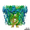 3524MC 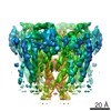 3523C 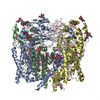 5mkeC M: map data used to model this data C: citing same article ( |
|---|---|
| Similar structure data |


-Links
 PDBj
PDBj


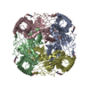
- Assembly
| Deposited unit | 
|
|---|---|
| 1 |
|
-Components
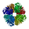
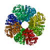
-Protein , 1 types, 4 molecules ABCD
| #1: Protein | Mass: 109820.086 Da / Num. of mol.: 4 Source method: isolated from a genetically manipulated source Source: (gene. exp.) Homo sapiens (human) / Gene: PKD2, TRPP2 / Cell line (production host): HEK293 / Production host: Homo sapiens (human) / References: UniProt: Q13563 |
|---|
-Sugars , 2 types, 16 molecules 
| #2: Polysaccharide | 2-acetamido-2-deoxy-beta-D-glucopyranose-(1-4)-2-acetamido-2-deoxy-beta-D-glucopyranose Source method: isolated from a genetically manipulated source #3: Sugar | ChemComp-NAG /  Type: D-saccharide, beta linking / Mass: 221.208 Da / Num. of mol.: 12 Type: D-saccharide, beta linking / Mass: 221.208 Da / Num. of mol.: 12Source method: isolated from a genetically manipulated source Formula: C8H15NO6 |
|---|
-Non-polymers , 4 types, 29 molecules 

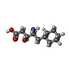

| #4: Chemical | ChemComp-PX6 /  Mass: 647.883 Da / Num. of mol.: 4 / Source method: obtained synthetically / Formula: C35H68O8P Mass: 647.883 Da / Num. of mol.: 4 / Source method: obtained synthetically / Formula: C35H68O8P#5: Chemical | ChemComp-PLM /  Mass: 256.424 Da / Num. of mol.: 12 / Source method: obtained synthetically / Formula: C16H32O2 Mass: 256.424 Da / Num. of mol.: 12 / Source method: obtained synthetically / Formula: C16H32O2#6: Chemical | ChemComp-CHS /  Type: peptide-like / Mass: 215.289 Da / Num. of mol.: 8 / Source method: obtained synthetically / Formula: C11H21NO3 Type: peptide-like / Mass: 215.289 Da / Num. of mol.: 8 / Source method: obtained synthetically / Formula: C11H21NO3#7: Chemical | ChemComp-CA /  Mass: 40.078 Da / Num. of mol.: 5 / Source method: obtained synthetically / Formula: Ca Mass: 40.078 Da / Num. of mol.: 5 / Source method: obtained synthetically / Formula: Ca |
|---|
-Details
| Has protein modification | Y |
|---|
-Experimental details
-Experiment
| Experiment | Method: ELECTRON MICROSCOPY |
|---|---|
| EM experiment | Aggregation state: PARTICLE / 3D reconstruction method: single particle reconstruction |
- Sample preparation
| Component | Name: Polycystin-2 / Type: COMPLEX / Entity ID: #1 / Source: RECOMBINANT |
|---|---|
| Source (natural) | Organism: Homo sapiens (human) |
| Source (recombinant) | Organism: Homo sapiens (human) |
| Buffer solution | pH: 7.5 |
| Specimen | Embedding applied: NO / Shadowing applied: NO / Staining applied: NO / Vitrification applied: YES |
| Vitrification | Cryogen name: ETHANE |
- Electron microscopy imaging
| Microscopy | Model: JEOL 3200FSC |
|---|---|
| Electron gun | Electron source:  FIELD EMISSION GUN / Accelerating voltage: 300 kV / Illumination mode: FLOOD BEAM FIELD EMISSION GUN / Accelerating voltage: 300 kV / Illumination mode: FLOOD BEAM |
| Electron lens | Mode: BRIGHT FIELD / Cs: 4.1 mm |
| Image recording | Electron dose: 1.8 e/Å2 / Film or detector model: GATAN K2 SUMMIT (4k x 4k) |
- Processing
| Software | Name: PHENIX / Version: 1.10.1_2155: / Classification: refinement | ||||||||||||||||||||||||
|---|---|---|---|---|---|---|---|---|---|---|---|---|---|---|---|---|---|---|---|---|---|---|---|---|---|
| EM software |
| ||||||||||||||||||||||||
| CTF correction | Type: PHASE FLIPPING AND AMPLITUDE CORRECTION | ||||||||||||||||||||||||
| Symmetry | Point symmetry: C4 (4 fold cyclic) | ||||||||||||||||||||||||
| 3D reconstruction | Resolution: 4.2 Å / Resolution method: FSC 0.143 CUT-OFF / Num. of particles: 35318 / Symmetry type: POINT | ||||||||||||||||||||||||
| Atomic model building | Space: REAL Details: we used for comparative structure modeling TRPA1 (pdb entry code 3J9P) as template for S1 and S3-S5, TRPV1 (pdb entry code 3J5Q) for S5-S6, and the TRPV2 (pdb entry code 5AN8) fitted best ...Details: we used for comparative structure modeling TRPA1 (pdb entry code 3J9P) as template for S1 and S3-S5, TRPV1 (pdb entry code 3J5Q) for S5-S6, and the TRPV2 (pdb entry code 5AN8) fitted best for S2-S3 to obtain an initial model. The soluble domain was build based on pdbID: 5K47. But we had no search model for molecular replacement. Although we had a good idea what the architecture would be like, we build the model de novo with COOT. | ||||||||||||||||||||||||
| Refine LS restraints |
|
