 Movie
Movie Controller

[English] 日本語
 Yorodumi
- PDB-6cxc: 3.9A Cryo-EM structure of murine antibody bound at a novel epitop... -
+ Open data

- Basic information
| Entry | Database: PDB / ID: 6cxc | ||||||||||||||||||||||||||||||||||||||||||||||||||||||
|---|---|---|---|---|---|---|---|---|---|---|---|---|---|---|---|---|---|---|---|---|---|---|---|---|---|---|---|---|---|---|---|---|---|---|---|---|---|---|---|---|---|---|---|---|---|---|---|---|---|---|---|---|---|---|---|
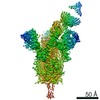
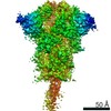
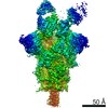
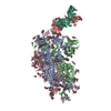
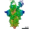
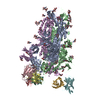
| Title | 3.9A Cryo-EM structure of murine antibody bound at a novel epitope of respiratory syncytial virus fusion protein | ||||||||||||||||||||||||||||||||||||||||||||||||||||||
 Components Components |
| ||||||||||||||||||||||||||||||||||||||||||||||||||||||
 Keywords Keywords | VIRAL PROTEIN/IMMUNE SYSTEM / respiratory syncytial virus fusion protein / murine antibody / novel epitope / complex / VIRAL PROTEIN-IMMUNE SYSTEM complex | ||||||||||||||||||||||||||||||||||||||||||||||||||||||
| Function / homology |  Function and homology information Function and homology informationsymbiont-mediated induction of syncytium formation / Translation of respiratory syncytial virus mRNAs / RSV-host interactions / Maturation of hRSV A proteins / Assembly and release of respiratory syncytial virus (RSV) virions / host cell Golgi membrane / Respiratory syncytial virus (RSV) attachment and entry / entry receptor-mediated virion attachment to host cell / fusion of virus membrane with host plasma membrane / viral envelope ...symbiont-mediated induction of syncytium formation / Translation of respiratory syncytial virus mRNAs / RSV-host interactions / Maturation of hRSV A proteins / Assembly and release of respiratory syncytial virus (RSV) virions / host cell Golgi membrane / Respiratory syncytial virus (RSV) attachment and entry / entry receptor-mediated virion attachment to host cell / fusion of virus membrane with host plasma membrane / viral envelope / symbiont entry into host cell / host cell plasma membrane / virion membrane / identical protein binding / plasma membrane Similarity search - Function | ||||||||||||||||||||||||||||||||||||||||||||||||||||||
| Biological species |   Human respiratory syncytial virus A Human respiratory syncytial virus A Human immunodeficiency virus 1 Human immunodeficiency virus 1 | ||||||||||||||||||||||||||||||||||||||||||||||||||||||
| Method | ELECTRON MICROSCOPY / single particle reconstruction / cryo EM / Resolution: 3.9 Å | ||||||||||||||||||||||||||||||||||||||||||||||||||||||
 Authors Authors | Xie, Q. / Wang, Z. / Chen, X. / Ni, F. / Ma, J. / Wang, Q. | ||||||||||||||||||||||||||||||||||||||||||||||||||||||
 Citation Citation | Journal: PLoS One / Year: 2019 Title: Structure basis of neutralization by a novel site II/IV antibody against respiratory syncytial virus fusion protein. Authors: Qingqing Xie / Zhao Wang / Fengyun Ni / Xiaorui Chen / Jianpeng Ma / Nita Patel / Hanxin Lu / Ye Liu / Jing-Hui Tian / David Flyer / Michael J Massare / Larry Ellingsworth / Gregory Glenn / ...Authors: Qingqing Xie / Zhao Wang / Fengyun Ni / Xiaorui Chen / Jianpeng Ma / Nita Patel / Hanxin Lu / Ye Liu / Jing-Hui Tian / David Flyer / Michael J Massare / Larry Ellingsworth / Gregory Glenn / Gale Smith / Qinghua Wang /  Abstract: Globally, human respiratory syncytial virus (RSV) is a leading cause of lower respiratory tract infections in newborns, young children, and the elderly for which there is no vaccine. The RSV fusion ...Globally, human respiratory syncytial virus (RSV) is a leading cause of lower respiratory tract infections in newborns, young children, and the elderly for which there is no vaccine. The RSV fusion (F) glycoprotein is a major target for vaccine development. Here, we describe a novel monoclonal antibody (designated as R4.C6) that recognizes both pre-fusion and post-fusion RSV F, and binds with nanomole affinity to a unique neutralizing site comprised of antigenic sites II and IV on the globular head. A 3.9 Å-resolution structure of RSV F-R4.C6 Fab complex was obtained by single particle cryo-electron microscopy and 3D reconstruction. The structure unraveled detailed interactions of R4.C6 with antigenic site II on one protomer and site IV on a neighboring protomer of post-fusion RSV F protein. These findings significantly further our understanding of the antigenic complexity of the F protein and provide new insights into RSV vaccine design. | ||||||||||||||||||||||||||||||||||||||||||||||||||||||
| History |
|
- Structure visualization
| Movie |
Movie viewer |
|---|---|
| Structure viewer | Molecule: MolmilJmol/JSmol |
- Downloads & links
-Download
| PDBx/mmCIF format | 6cxc.cif.gz | 372.9 KB | Display | PDBx/mmCIF format |
|---|---|---|---|---|
| PDB format | pdb6cxc.ent.gz | 287.7 KB | Display | PDB format |
| PDBx/mmJSON format | 6cxc.json.gz | Tree view | PDBx/mmJSON format | |
| Others |  Other downloads Other downloads |
-Validation report
| Summary document | 6cxc_validation.pdf.gz | 1 MB | Display | wwPDB validaton report |
|---|---|---|---|---|
| Full document | 6cxc_full_validation.pdf.gz | 1.1 MB | Display | |
| Data in XML | 6cxc_validation.xml.gz | 60.7 KB | Display | |
| Data in CIF | 6cxc_validation.cif.gz | 90.8 KB | Display | |
| Arichive directory | https://data.pdbj.org/pub/pdb/validation_reports/cx/6cxcftp://data.pdbj.org/pub/pdb/validation_reports/cx/6cxc | HTTPS FTP |
-Related structure data
| Related structure data |  7774MC M: map data used to model this data C: citing same article ( |
|---|---|
| Similar structure data |
-Links
 PDBj
PDBj


- Assembly
| Deposited unit | 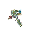
|
|---|---|
| 1 |
|
-Components
| #1: Antibody | Mass: 12831.376 Da / Num. of mol.: 3 Source method: isolated from a genetically manipulated source Source: (gene. exp.) #2: Antibody | Mass: 13457.151 Da / Num. of mol.: 3 Source method: isolated from a genetically manipulated source Source: (gene. exp.) #3: Protein | Mass: 60728.047 Da / Num. of mol.: 6 Source method: isolated from a genetically manipulated source Source: (gene. exp.) Human respiratory syncytial virus A (strain A2), (gene. exp.) Human immunodeficiency virus 1Strain: A2 / Production host:   Spodoptera frugiperda (fall armyworm) / References: UniProt: P03420, UniProt: M1E1E4 Spodoptera frugiperda (fall armyworm) / References: UniProt: P03420, UniProt: M1E1E4#4: Sugar | ChemComp-NAG /   Type: D-saccharide, beta linking / Mass: 221.208 Da / Num. of mol.: 6 Type: D-saccharide, beta linking / Mass: 221.208 Da / Num. of mol.: 6Source method: isolated from a genetically manipulated source Formula: C8H15NO6 Has ligand of interest | N | Has protein modification | Y | |
|---|
-Experimental details
-Experiment
| Experiment | Method: ELECTRON MICROSCOPY |
|---|---|
| EM experiment | Aggregation state: PARTICLE / 3D reconstruction method: single particle reconstruction |
- Sample preparation
| Component |
| ||||||||||||||||||||||||
|---|---|---|---|---|---|---|---|---|---|---|---|---|---|---|---|---|---|---|---|---|---|---|---|---|---|
| Source (natural) |
| ||||||||||||||||||||||||
| Source (recombinant) |
| ||||||||||||||||||||||||
| Buffer solution | pH: 7 | ||||||||||||||||||||||||
| Specimen | Embedding applied: NO / Shadowing applied: NO / Staining applied: NO / Vitrification applied: YES | ||||||||||||||||||||||||
| Specimen support | Grid material: COPPER / Grid mesh size: 200 divisions/in. / Grid type: Quantifoil R1.2/1.3 | ||||||||||||||||||||||||
| Vitrification | Instrument: LEICA EM GP / Cryogen name: NITROGEN / Humidity: 98 % / Chamber temperature: 295 K |
- Electron microscopy imaging
| Microscopy | Model: JEOL 3200FSC |
|---|---|
| Electron gun | Electron source:  FIELD EMISSION GUN / Accelerating voltage: 300 kV / Illumination mode: FLOOD BEAM FIELD EMISSION GUN / Accelerating voltage: 300 kV / Illumination mode: FLOOD BEAM |
| Electron lens | Mode: BRIGHT FIELD / Cs: 4.7 mm / C2 aperture diameter: 50 µm |
| Specimen holder | Cryogen: NITROGEN / Specimen holder model: JEOL 3200FSC CRYOHOLDER |
| Image recording | Average exposure time: 10 sec. / Electron dose: 50 e/Å2 / Detector mode: SUPER-RESOLUTION / Film or detector model: GATAN K2 SUMMIT (4k x 4k) |
| Image scans | Movie frames/image: 50 |
- Processing
| Software | Name: PHENIX / Version: 1.11.1_2575: / Classification: refinement | ||||||||||||||||||||||||||||||||
|---|---|---|---|---|---|---|---|---|---|---|---|---|---|---|---|---|---|---|---|---|---|---|---|---|---|---|---|---|---|---|---|---|---|
| EM software |
| ||||||||||||||||||||||||||||||||
| CTF correction | Type: PHASE FLIPPING AND AMPLITUDE CORRECTION | ||||||||||||||||||||||||||||||||
| Symmetry | Point symmetry: C3 (3 fold cyclic) | ||||||||||||||||||||||||||||||||
| 3D reconstruction | Resolution: 3.9 Å / Resolution method: FSC 0.143 CUT-OFF / Num. of particles: 543639 / Symmetry type: POINT | ||||||||||||||||||||||||||||||||
| Atomic model building | Protocol: RIGID BODY FIT / Space: REAL | ||||||||||||||||||||||||||||||||
| Refine LS restraints |
|
