 Movie
Movie Controller

[English] 日本語
 Yorodumi
- EMDB-7774: 3.9A Cryo-EM structure of murine antibody bound at a novel epitop... -
+ Open data

- Basic information
| Entry | Database: EMDB / ID: EMD-7774 | |||||||||
|---|---|---|---|---|---|---|---|---|---|---|
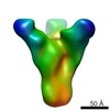
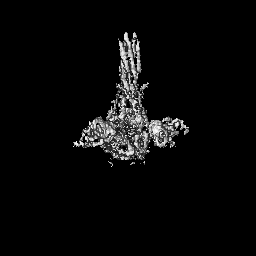
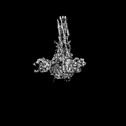
| Title | 3.9A Cryo-EM structure of murine antibody bound at a novel epitope of respiratory syncytial virus fusion protein | |||||||||
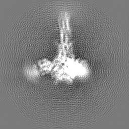
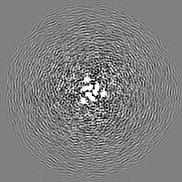
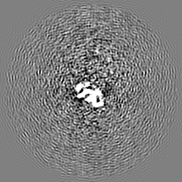
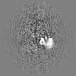
 Map data Map data | 3.9A density map of R4.C6 in complex with RSV F710 | |||||||||
 Sample Sample |
| |||||||||
 Keywords Keywords | respiratory syncytial virus fusion protein / murine antibody / novel epitope / complex / VIRAL PROTEIN-IMMUNE SYSTEM complex | |||||||||
| Function / homology |  Function and homology information Function and homology informationsymbiont-mediated induction of syncytium formation / Translation of respiratory syncytial virus mRNAs / RSV-host interactions / Assembly and release of respiratory syncytial virus (RSV) virions / Maturation of hRSV A proteins / Respiratory syncytial virus (RSV) attachment and entry / host cell Golgi membrane / entry receptor-mediated virion attachment to host cell / fusion of virus membrane with host plasma membrane / viral envelope ...symbiont-mediated induction of syncytium formation / Translation of respiratory syncytial virus mRNAs / RSV-host interactions / Assembly and release of respiratory syncytial virus (RSV) virions / Maturation of hRSV A proteins / Respiratory syncytial virus (RSV) attachment and entry / host cell Golgi membrane / entry receptor-mediated virion attachment to host cell / fusion of virus membrane with host plasma membrane / viral envelope / symbiont entry into host cell / host cell plasma membrane / virion membrane / identical protein binding / plasma membrane Similarity search - Function | |||||||||
| Biological species |   Human respiratory syncytial virus A2 / Human respiratory syncytial virus A2 /  Human immunodeficiency virus 1 Human immunodeficiency virus 1 | |||||||||
| Method | single particle reconstruction / cryo EM / Resolution: 3.9 Å | |||||||||
 Authors Authors | Xie Q / Wang Z | |||||||||
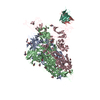
 Citation Citation | Journal: PLoS One / Year: 2019 Title: Structure basis of neutralization by a novel site II/IV antibody against respiratory syncytial virus fusion protein. Authors: Qingqing Xie / Zhao Wang / Fengyun Ni / Xiaorui Chen / Jianpeng Ma / Nita Patel / Hanxin Lu / Ye Liu / Jing-Hui Tian / David Flyer / Michael J Massare / Larry Ellingsworth / Gregory Glenn / ...Authors: Qingqing Xie / Zhao Wang / Fengyun Ni / Xiaorui Chen / Jianpeng Ma / Nita Patel / Hanxin Lu / Ye Liu / Jing-Hui Tian / David Flyer / Michael J Massare / Larry Ellingsworth / Gregory Glenn / Gale Smith / Qinghua Wang /  Abstract: Globally, human respiratory syncytial virus (RSV) is a leading cause of lower respiratory tract infections in newborns, young children, and the elderly for which there is no vaccine. The RSV fusion ...Globally, human respiratory syncytial virus (RSV) is a leading cause of lower respiratory tract infections in newborns, young children, and the elderly for which there is no vaccine. The RSV fusion (F) glycoprotein is a major target for vaccine development. Here, we describe a novel monoclonal antibody (designated as R4.C6) that recognizes both pre-fusion and post-fusion RSV F, and binds with nanomole affinity to a unique neutralizing site comprised of antigenic sites II and IV on the globular head. A 3.9 Å-resolution structure of RSV F-R4.C6 Fab complex was obtained by single particle cryo-electron microscopy and 3D reconstruction. The structure unraveled detailed interactions of R4.C6 with antigenic site II on one protomer and site IV on a neighboring protomer of post-fusion RSV F protein. These findings significantly further our understanding of the antigenic complexity of the F protein and provide new insights into RSV vaccine design. | |||||||||
| History |
|
- Structure visualization
| Movie |
Movie viewer |
|---|---|
| Structure viewer | EM map: SurfViewMolmilJmol/JSmol |
| Supplemental images |

- Downloads & links
-EMDB archive
| Map data | emd_7774.map.gz | 59.9 MB | EMDB map data format | |
|---|---|---|---|---|
| Header (meta data) | emd-7774-v30.xmlemd-7774.xml | 20.3 KB 20.3 KB | Display Display | EMDB header |
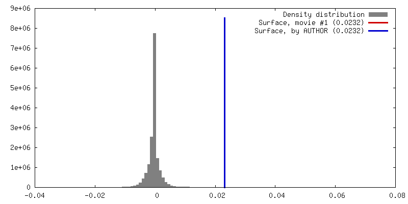
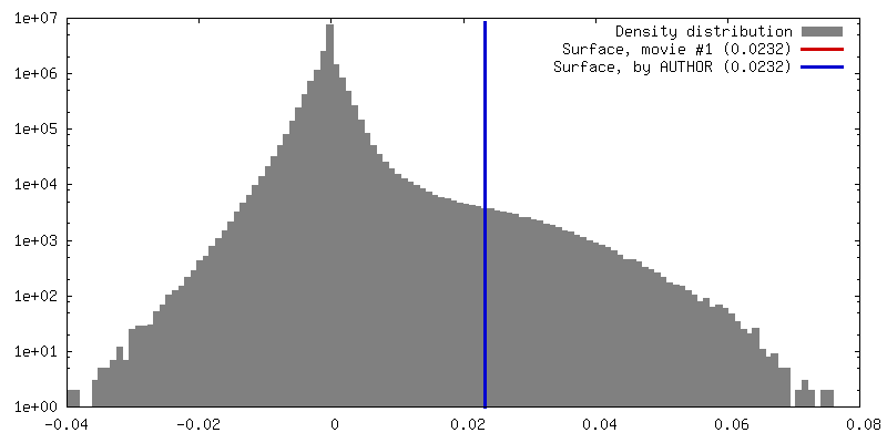
| FSC (resolution estimation) | emd_7774_fsc.xml | 9.1 KB | Display | FSC data file |
| Images | 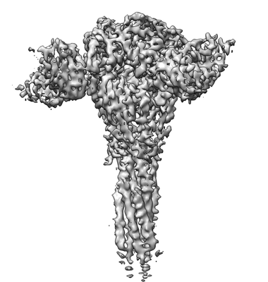 emd_7774.png emd_7774.png | 47.2 KB | ||
| Filedesc metadata | emd-7774.cif.gz | 7.2 KB | ||
| Archive directory |  http://ftp.pdbj.org/pub/emdb/structures/EMD-7774ftp://ftp.pdbj.org/pub/emdb/structures/EMD-7774 http://ftp.pdbj.org/pub/emdb/structures/EMD-7774ftp://ftp.pdbj.org/pub/emdb/structures/EMD-7774 | HTTPS FTP |
-Related structure data
| Related structure data |  6cxcMC M: atomic model generated by this map C: citing same article ( |
|---|---|
| Similar structure data |


-Links
| EMDB pages | EMDB (EBI/PDBe) / EMDataResource |
|---|---|
| Related items in Molecule of the Month |


-Map
| File | Download / File: emd_7774.map.gz / Format: CCP4 / Size: 64 MB / Type: IMAGE STORED AS FLOATING POINT NUMBER (4 BYTES) | ||||||||||||||||||||||||||||||||||||||||||||||||||||||||||||||||||||
|---|---|---|---|---|---|---|---|---|---|---|---|---|---|---|---|---|---|---|---|---|---|---|---|---|---|---|---|---|---|---|---|---|---|---|---|---|---|---|---|---|---|---|---|---|---|---|---|---|---|---|---|---|---|---|---|---|---|---|---|---|---|---|---|---|---|---|---|---|---|
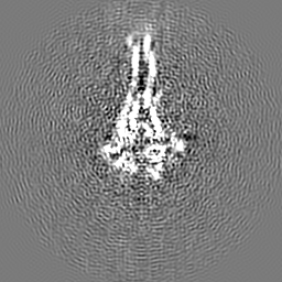
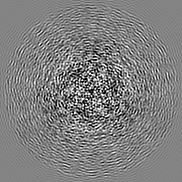
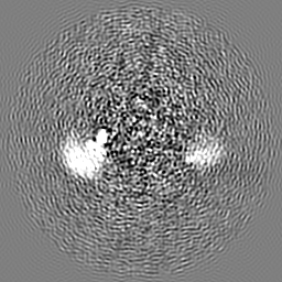
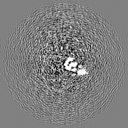
| Annotation | 3.9A density map of R4.C6 in complex with RSV F710 | ||||||||||||||||||||||||||||||||||||||||||||||||||||||||||||||||||||
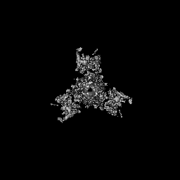
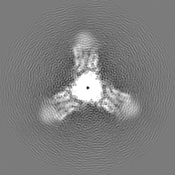
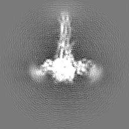
| Projections & slices | Image control
Images are generated by Spider. | ||||||||||||||||||||||||||||||||||||||||||||||||||||||||||||||||||||
| Voxel size | X=Y=Z: 1.2546 Å | ||||||||||||||||||||||||||||||||||||||||||||||||||||||||||||||||||||
| Density |
| ||||||||||||||||||||||||||||||||||||||||||||||||||||||||||||||||||||
| Symmetry | Space group: 1 | ||||||||||||||||||||||||||||||||||||||||||||||||||||||||||||||||||||
| Details | EMDB XML:
CCP4 map header:
| ||||||||||||||||||||||||||||||||||||||||||||||||||||||||||||||||||||
 Z (Sec.)
 Y (Row.)
 X (Col.)


-Supplemental data
- Sample components
-Entire : Complex of R4.C6 Fab with RSV F protein
| Entire | Name: Complex of R4.C6 Fab with RSV F protein |
|---|---|
| Components |
|
-Supramolecule #1: Complex of R4.C6 Fab with RSV F protein
| Supramolecule | Name: Complex of R4.C6 Fab with RSV F protein / type: complex / ID: 1 / Parent: 0 / Macromolecule list: #1-#3 |
|---|
-Supramolecule #2: R4.C6 Fab
| Supramolecule | Name: R4.C6 Fab / type: complex / ID: 2 / Parent: 1 / Macromolecule list: #1-#2 |
|---|---|
| Source (natural) | Organism: |
-Supramolecule #3: RSV F protein
| Supramolecule | Name: RSV F protein / type: complex / ID: 3 / Parent: 1 / Macromolecule list: #3 |
|---|---|
| Source (natural) | Organism: Human respiratory syncytial virus A2 |
-Macromolecule #1: R4.C6 Fab Heavy Chain
| Macromolecule | Name: R4.C6 Fab Heavy Chain / type: protein_or_peptide / ID: 1 / Number of copies: 3 / Enantiomer: LEVO |
|---|---|
| Source (natural) | Organism: |
| Molecular weight | Theoretical: 12.831376 KDa |
| Recombinant expression | Organism: |
| Sequence | String: MAEVQLQQSG PELVKPGASV KISCKASGYA FSNSWMSWVK QRPGKGLEWI GRLFPADGDI TYNGHFKDKA ALTADKSSNT AYIQLSSLT SEDSAVYFCA RMDNSEVFWG QGTLVTVSA |
-Macromolecule #2: R4.C6 Fab Light Chain
| Macromolecule | Name: R4.C6 Fab Light Chain / type: protein_or_peptide / ID: 2 / Number of copies: 3 / Enantiomer: LEVO |
|---|---|
| Source (natural) | Organism: |
| Molecular weight | Theoretical: 13.457151 KDa |
| Recombinant expression | Organism: |
| Sequence | String: MADILLTQSQ KFMSTSVGDR VSITCKASQN VRTGVSWYQR KPGQSPKALI YLASNRHTGV PDRFTGRGSG TDFTLTISEV QSEDLADYF CLQHWTVPYT FGGGTKLEIK RTAAAPSRAN SFK |
-Macromolecule #3: Fusion glycoprotein F0, Envelope glycoprotein chimera
| Macromolecule | Name: Fusion glycoprotein F0, Envelope glycoprotein chimera / type: protein_or_peptide / ID: 3 / Number of copies: 6 / Enantiomer: LEVO |
|---|---|
| Source (natural) | Organism: Human immunodeficiency virus 1 |
| Molecular weight | Theoretical: 60.728047 KDa |
| Recombinant expression | Organism:   Spodoptera frugiperda (fall armyworm) Spodoptera frugiperda (fall armyworm) |
| Sequence | String: QNITEEFYQS TCSAVSKGYL SALRTGWYTS VITIELSNIK ENKCNGTDAK VKLIKQELDK YKNAVTELQL LMQSTPATNN RARRELPRF MNYTLNNAKK TNVTLSKKQK QQFLGFLLGV GSAIASGVAV SKVLHLEGEV NKIKSALLST NKAVVSLSNG V SVLTSKVL ...String: QNITEEFYQS TCSAVSKGYL SALRTGWYTS VITIELSNIK ENKCNGTDAK VKLIKQELDK YKNAVTELQL LMQSTPATNN RARRELPRF MNYTLNNAKK TNVTLSKKQK QQFLGFLLGV GSAIASGVAV SKVLHLEGEV NKIKSALLST NKAVVSLSNG V SVLTSKVL DLKNYIDKQL LPIVNKQSCS ISNIETVIEF QQKNNRLLEI TREFSVNAGV TTPVSTYMLT NSELLSLIND MP ITNDQKK LMSNNVQIVR QQSYSIMSII KEEVLAYVVQ LPLYGVIDTP CWKLHTSPLC TTNTKEGSNI CLTRTDRGWY CDN AGSVSF FPQAETCKVQ SNRVFCDTMN SLTLPSEVNL CNVDIFNPKY DCKIMTSKTD VSSSVITSLG AIVSCYGKTK CTAS NKNRG IIKTFSNGCD YVSNKGVDTV SVGNTLYYVN KQEGKSLYVK GEPIINFYDP LVFPSDEFDA SISQVNEKIN QSLAF IRKS DELLHNVNAG KSTTNIMGAL VPRGSPGSGY IPEAPRDGQA YVRKDGEWVL LSTFLGSSHH HHHH UniProtKB: Fusion glycoprotein F0, Envelope glycoprotein |
-Macromolecule #4: 2-acetamido-2-deoxy-beta-D-glucopyranose
| Macromolecule | Name: 2-acetamido-2-deoxy-beta-D-glucopyranose / type: ligand / ID: 4 / Number of copies: 6 / Formula: NAG |
|---|---|
| Molecular weight | Theoretical: 221.208 Da |
| Chemical component information |  ChemComp-NAG: |
-Experimental details
-Structure determination
| Method | cryo EM |
|---|---|
 Processing Processing | single particle reconstruction |
| Aggregation state | particle |
-Sample preparation
| Buffer | pH: 7 |
|---|---|
| Grid | Model: Quantifoil R1.2/1.3 / Material: COPPER / Mesh: 200 / Support film - Material: GRAPHENE OXIDE |
| Vitrification | Cryogen name: NITROGEN / Chamber humidity: 98 % / Chamber temperature: 295 K / Instrument: LEICA EM GP |
- Electron microscopy
| Microscope | JEOL 3200FSC |
|---|---|
| Image recording | Film or detector model: GATAN K2 SUMMIT (4k x 4k) / Detector mode: SUPER-RESOLUTION / Average exposure time: 10.0 sec. / Average electron dose: 50.0 e/Å2 |
| Electron beam | Acceleration voltage: 300 kV / Electron source:  FIELD EMISSION GUN FIELD EMISSION GUN |
| Electron optics | C2 aperture diameter: 50.0 µm / Illumination mode: FLOOD BEAM / Imaging mode: BRIGHT FIELD / Cs: 4.7 mm |
| Sample stage | Specimen holder model: JEOL 3200FSC CRYOHOLDER / Cooling holder cryogen: NITROGEN |
+Image processing

-Atomic model buiding 1
| Refinement | Space: REAL / Protocol: RIGID BODY FIT |
|---|---|
| Output model | PDB-6cxc: |
