 Movie
Movie Controller

[English] 日本語
 Yorodumi
- PDB-6wf7: Methylmalonyl-CoA epimerase in complex with methylmalonyl-CoA and NH4+ -
+ Open data

- Basic information
| Entry | Database: PDB / ID: 6wf7 | ||||||
|---|---|---|---|---|---|---|---|
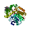
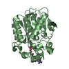
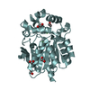
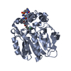
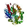
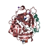
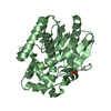
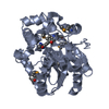
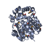
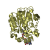
| Title | Methylmalonyl-CoA epimerase in complex with methylmalonyl-CoA and NH4+ | ||||||
 Components Components | Methylmalonyl-CoA epimerase | ||||||
 Keywords Keywords | ISOMERASE / Epimerase / acid-base / enol / enolate | ||||||
| Function / homology |  Function and homology information Function and homology informationmethylmalonyl-CoA epimerase activity / L-methylmalonyl-CoA metabolic process / metal ion binding Similarity search - Function | ||||||
| Biological species |  Streptomyces coelicolor (bacteria) Streptomyces coelicolor (bacteria) | ||||||
| Method |  X-RAY DIFFRACTION / SYNCHROTRON / MOLECULAR REPLACEMENT / Resolution: 1.55 Å X-RAY DIFFRACTION / SYNCHROTRON / MOLECULAR REPLACEMENT / Resolution: 1.55 Å | ||||||
 Authors Authors | Stunkard, L.M. / Benjamin, A.B. / Bower, J.B. / Huth, T.J. / Lohman, J.R. | ||||||
 Citation Citation | Journal: Chembiochem / Year: 2022 Title: Substrate Enolate Intermediate and Mimic Captured in the Active Site of Streptomyces coelicolor Methylmalonyl-CoA Epimerase. Authors: Stunkard, L.M. / Benjamin, A.B. / Bower, J.B. / Huth, T.J. / Lohman, J.R. | ||||||
| History |
|
- Structure visualization
| Structure viewer | Molecule: MolmilJmol/JSmol |
|---|
- Downloads & links
-Download
| PDBx/mmCIF format | 6wf7.cif.gz | 56.7 KB | Display | PDBx/mmCIF format |
|---|---|---|---|---|
| PDB format | pdb6wf7.ent.gz | 36.7 KB | Display | PDB format |
| PDBx/mmJSON format | 6wf7.json.gz | Tree view | PDBx/mmJSON format | |
| Others |  Other downloads Other downloads |
-Validation report
| Arichive directory | https://data.pdbj.org/pub/pdb/validation_reports/wf/6wf7ftp://data.pdbj.org/pub/pdb/validation_reports/wf/6wf7 | HTTPS FTP |
|---|
-Related structure data
| Related structure data |  6wf6C 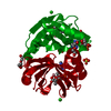 6wfhC 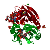 6wfiC  1jc5S S: Starting model for refinement C: citing same article ( |
|---|---|
| Similar structure data |
-Links
 PDBj
PDBj
- Assembly
| Deposited unit | 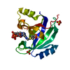
| |||||||||
|---|---|---|---|---|---|---|---|---|---|---|
| 1 | 
| |||||||||
| Unit cell |
| |||||||||
| Components on special symmetry positions |
|
-Components
-Protein , 1 types, 1 molecules A
| #1: Protein | Mass: 16059.767 Da / Num. of mol.: 1 / Mutation: M1S Source method: isolated from a genetically manipulated source Source: (gene. exp.) Streptomyces coelicolor (bacteria) / Strain: ATCC BAA-471 / A3(2) / M145 / Gene: SCO5398 / Production host: |
|---|
-Non-polymers , 5 types, 207 molecules 


| #2: Chemical | ChemComp-NH4 /  Mass: 18.038 Da / Num. of mol.: 1 / Source method: obtained synthetically / Formula: H4N Mass: 18.038 Da / Num. of mol.: 1 / Source method: obtained synthetically / Formula: H4N | ||||||
|---|---|---|---|---|---|---|---|
| #3: Chemical |  Mass: 96.063 Da / Num. of mol.: 2 / Source method: obtained synthetically / Formula: SO4 Mass: 96.063 Da / Num. of mol.: 2 / Source method: obtained synthetically / Formula: SO4#4: Chemical | ChemComp-MC0 / ( |  Mass: 867.607 Da / Num. of mol.: 1 / Source method: obtained synthetically / Formula: C25H40N7O19P3S / Feature type: SUBJECT OF INVESTIGATION Mass: 867.607 Da / Num. of mol.: 1 / Source method: obtained synthetically / Formula: C25H40N7O19P3S / Feature type: SUBJECT OF INVESTIGATION#5: Chemical | ChemComp-MCA / |  Mass: 867.607 Da / Num. of mol.: 1 / Source method: obtained synthetically / Formula: C25H40N7O19P3S / Feature type: SUBJECT OF INVESTIGATION Mass: 867.607 Da / Num. of mol.: 1 / Source method: obtained synthetically / Formula: C25H40N7O19P3S / Feature type: SUBJECT OF INVESTIGATION#6: Water | ChemComp-HOH / | Mass: 18.015 Da / Num. of mol.: 202 / Source method: isolated from a natural source / Formula: H2O |
-Details
| Has ligand of interest | Y |
|---|
-Experimental details
-Experiment
| Experiment | Method: X-RAY DIFFRACTION / Number of used crystals: 1 |
|---|
- Sample preparation
| Crystal | Density Matthews: 3.69 Å3/Da / Density % sol: 66.66 % |
|---|---|
| Crystal grow | Temperature: 298 K / Method: vapor diffusion, hanging drop / pH: 7 Details: 50 mM sodium chloride, 100 mM Bis-Tris:HCl pH 7.0, 2.3 M ammonium sulfate, 5% PEG 400 |
-Data collection
| Diffraction | Mean temperature: 80 K / Ambient temp details: liquid nitrogen / Serial crystal experiment: N |
|---|---|
| Diffraction source | Source: SYNCHROTRON / Site: APS  / Beamline: 21-ID-F / Wavelength: 0.97872 Å / Beamline: 21-ID-F / Wavelength: 0.97872 Å |
| Detector | Type: RAYONIX MX300HE / Detector: CCD / Date: Jul 16, 2017 / Details: MD2 Micro Diffractometer |
| Diffraction measurement | Details: 1.00 degrees, 3.06 sec, detector distance 200.00 mm Method: \w scans |
| Radiation | Monochromator: C(111) / Protocol: SINGLE WAVELENGTH / Monochromatic (M) / Laue (L): M / Scattering type: x-ray |
| Radiation wavelength | Wavelength: 0.97872 Å / Relative weight: 1 |
| Reflection | Av R equivalents: 0.087 / Number: 521570 |
| Reflection | Resolution: 1.55→30 Å / Num. obs: 36333 / % possible obs: 100 % / Observed criterion σ(F): 0 / Observed criterion σ(I): -3 / Redundancy: 14.4 % / Rmerge(I) obs: 0.087 / Rpim(I) all: 0.024 / Rrim(I) all: 0.09 / Rsym value: 0.087 / Χ2: 1.015 / Net I/av σ(I): 27 / Net I/σ(I): 11.4 / Num. measured all: 521570 |
| Reflection shell | Resolution: 1.55→1.61 Å / Redundancy: 14.1 % / Rmerge(I) obs: 0.614 / Mean I/σ(I) obs: 5.5 / Num. unique obs: 3564 / Rsym value: 0.614 / % possible all: 100 |
| Cell measurement | Reflection used: 521570 |
- Processing
| Software |
| ||||||||||||||||||||||||||||||||||||||||||||||||||||||||||||
|---|---|---|---|---|---|---|---|---|---|---|---|---|---|---|---|---|---|---|---|---|---|---|---|---|---|---|---|---|---|---|---|---|---|---|---|---|---|---|---|---|---|---|---|---|---|---|---|---|---|---|---|---|---|---|---|---|---|---|---|---|---|
| Refinement | Method to determine structure: MOLECULAR REPLACEMENT Starting model: 1JC5 Resolution: 1.55→29.4 Å / Cor.coef. Fo:Fc: 0.975 / Cor.coef. Fo:Fc free: 0.969 / WRfactor Rfree: 0.1951 / WRfactor Rwork: 0.1803 / FOM work R set: 0.9022 / SU B: 0.84 / SU ML: 0.031 / SU R Cruickshank DPI: 0.0539 / SU Rfree: 0.0552 / Cross valid method: THROUGHOUT / σ(F): 0 / ESU R: 0.054 / ESU R Free: 0.055 / Stereochemistry target values: MAXIMUM LIKELIHOOD Details: HYDROGENS HAVE BEEN ADDED IN THE RIDING POSITIONS U VALUES : REFINED INDIVIDUALLY
| ||||||||||||||||||||||||||||||||||||||||||||||||||||||||||||
| Solvent computation | Ion probe radii: 0.8 Å / Shrinkage radii: 0.8 Å / VDW probe radii: 1.2 Å / Solvent model: MASK | ||||||||||||||||||||||||||||||||||||||||||||||||||||||||||||
| Displacement parameters | Biso max: 799 Å2 / Biso mean: 24.875 Å2 / Biso min: 12.92 Å2
| ||||||||||||||||||||||||||||||||||||||||||||||||||||||||||||
| Refinement step | Cycle: final / Resolution: 1.55→29.4 Å
| ||||||||||||||||||||||||||||||||||||||||||||||||||||||||||||
| Refine LS restraints |
| ||||||||||||||||||||||||||||||||||||||||||||||||||||||||||||
| LS refinement shell | Resolution: 1.55→1.591 Å / Rfactor Rfree error: 0 / Total num. of bins used: 20
|
