 Movie
Movie Controller

[English] 日本語
 Yorodumi
- PDB-6ufp: Structure of proline utilization A with the FAD covalently modifi... -
+ Open data

- Basic information
| Entry | Database: PDB / ID: 6ufp | |||||||||
|---|---|---|---|---|---|---|---|---|---|---|
| Title | Structure of proline utilization A with the FAD covalently modified by L-thiazolidine-2-carboxylate and three cysteines (Cys46, Cys470, Cys638) modified to S,S-(2-HYDROXYETHYL)THIOCYSTEINE | |||||||||
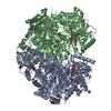
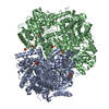
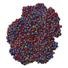
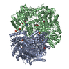
 Components Components | (Bifunctional protein ...) x 2 | |||||||||
 Keywords Keywords | OXIDOREDUCTASE / FLAVOENZYME / ROSSMANN FOLD / PROLINE CATABOLISM / SUBSTRATE CHANNELING / BIFUNCTIONAL ENZYME | |||||||||
| Function / homology |  Function and homology information Function and homology informationproline dehydrogenase / proline dehydrogenase activity / L-glutamate gamma-semialdehyde dehydrogenase / L-glutamate gamma-semialdehyde dehydrogenase activity / : / cytoplasmic side of plasma membrane / DNA-binding transcription factor activity / nucleotide binding / DNA binding Similarity search - Function | |||||||||
| Biological species |  Sinorhizobium meliloti (bacteria) Sinorhizobium meliloti (bacteria) | |||||||||
| Method |  X-RAY DIFFRACTION / SYNCHROTRON / FOURIER SYNTHESIS / Resolution: 1.737 Å X-RAY DIFFRACTION / SYNCHROTRON / FOURIER SYNTHESIS / Resolution: 1.737 Å | |||||||||
 Authors Authors | Campbell, A.C. / Tanner, J.J. | |||||||||
| Funding support |  United States, 2items United States, 2items
| |||||||||
 Citation Citation | Journal: Acs Chem.Biol. / Year: 2020 Title: Covalent Modification of the Flavin in Proline Dehydrogenase by Thiazolidine-2-Carboxylate. Authors: Campbell, A.C. / Becker, D.F. / Gates, K.S. / Tanner, J.J. | |||||||||
| History |
|
- Structure visualization
| Structure viewer | Molecule: MolmilJmol/JSmol |
|---|
- Downloads & links
-Download
| PDBx/mmCIF format | 6ufp.cif.gz | 924.2 KB | Display | PDBx/mmCIF format |
|---|---|---|---|---|
| PDB format | pdb6ufp.ent.gz | 748.1 KB | Display | PDB format |
| PDBx/mmJSON format | 6ufp.json.gz | Tree view | PDBx/mmJSON format | |
| Others |  Other downloads Other downloads |
-Validation report
| Arichive directory | https://data.pdbj.org/pub/pdb/validation_reports/uf/6ufpftp://data.pdbj.org/pub/pdb/validation_reports/uf/6ufp | HTTPS FTP |
|---|
-Related structure data
| Related structure data |  6vz9C 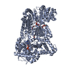 5kf6S S: Starting model for refinement C: citing same article ( |
|---|---|
| Similar structure data |


-Links
 PDBj
PDBj

- Assembly
| Deposited unit | 
| ||||||||
|---|---|---|---|---|---|---|---|---|---|
| 1 |
| ||||||||
| Unit cell |
|
-Components
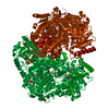
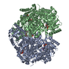
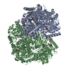
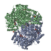
-Bifunctional protein ... , 2 types, 2 molecules AB
| #1: Protein | Mass: 132113.891 Da / Num. of mol.: 1 Source method: isolated from a genetically manipulated source Source: (gene. exp.) Sinorhizobium meliloti (strain SM11) (bacteria)Strain: SM11 / Gene: putA, SM11_chr0102 / Production host: References: UniProt: F7X6I3, proline dehydrogenase, L-glutamate gamma-semialdehyde dehydrogenase |
|---|---|
| #2: Protein | Mass: 132190.000 Da / Num. of mol.: 1 Source method: isolated from a genetically manipulated source Source: (gene. exp.) Sinorhizobium meliloti (strain SM11) (bacteria)Strain: SM11 / Gene: putA, SM11_chr0102 / Production host: References: UniProt: F7X6I3, proline dehydrogenase, L-glutamate gamma-semialdehyde dehydrogenase |
-Non-polymers , 7 types, 1235 molecules 
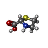


| #3: Chemical |  Mass: 787.566 Da / Num. of mol.: 2 / Source method: obtained synthetically / Formula: C27H35N9O15P2 / Feature type: SUBJECT OF INVESTIGATION Mass: 787.566 Da / Num. of mol.: 2 / Source method: obtained synthetically / Formula: C27H35N9O15P2 / Feature type: SUBJECT OF INVESTIGATION#4: Chemical |  Mass: 133.169 Da / Num. of mol.: 2 / Source method: obtained synthetically / Formula: C4H7NO2S / Feature type: SUBJECT OF INVESTIGATION Mass: 133.169 Da / Num. of mol.: 2 / Source method: obtained synthetically / Formula: C4H7NO2S / Feature type: SUBJECT OF INVESTIGATION#5: Chemical |  Mass: 663.425 Da / Num. of mol.: 2 / Source method: obtained synthetically / Formula: C21H27N7O14P2 / Feature type: SUBJECT OF INVESTIGATION / Comment: NAD*YM Mass: 663.425 Da / Num. of mol.: 2 / Source method: obtained synthetically / Formula: C21H27N7O14P2 / Feature type: SUBJECT OF INVESTIGATION / Comment: NAD*YM#6: Chemical |  Mass: 106.120 Da / Num. of mol.: 2 / Source method: obtained synthetically / Formula: C4H10O3 Mass: 106.120 Da / Num. of mol.: 2 / Source method: obtained synthetically / Formula: C4H10O3#7: Chemical | ChemComp-SO4 /  Mass: 96.063 Da / Num. of mol.: 6 / Source method: obtained synthetically / Formula: SO4 Mass: 96.063 Da / Num. of mol.: 6 / Source method: obtained synthetically / Formula: SO4#8: Chemical |  Mass: 46.025 Da / Num. of mol.: 2 / Source method: obtained synthetically / Formula: CH2O2 Mass: 46.025 Da / Num. of mol.: 2 / Source method: obtained synthetically / Formula: CH2O2#9: Water | ChemComp-HOH / | Mass: 18.015 Da / Num. of mol.: 1219 / Source method: isolated from a natural source / Formula: H2O |
|---|
-Details
| Has ligand of interest | Y |
|---|---|
| Has protein modification | Y |
-Experimental details
-Experiment
| Experiment | Method: X-RAY DIFFRACTION / Number of used crystals: 1 |
|---|
- Sample preparation
| Crystal | Density Matthews: 2.38 Å3/Da / Density % sol: 48.4 % |
|---|---|
| Crystal grow | Temperature: 286 K / Method: vapor diffusion, sitting drop / pH: 8 Details: 20% PEG-3350, 0.2M ammonium sulfate, 0.1M MgCl2, 0.1M HEPES, 0.1M Na formate, 10mM NAD+, 50mM T2C |
-Data collection
| Diffraction | Mean temperature: 100 K / Serial crystal experiment: N | ||||||||||||||||||||||||||||||
|---|---|---|---|---|---|---|---|---|---|---|---|---|---|---|---|---|---|---|---|---|---|---|---|---|---|---|---|---|---|---|---|
| Diffraction source | Source: SYNCHROTRON / Site: APS / Beamline: 24-ID-C / Wavelength: 0.9791 Å | ||||||||||||||||||||||||||||||
| Detector | Type: DECTRIS PILATUS3 6M / Detector: PIXEL / Date: Aug 3, 2018 | ||||||||||||||||||||||||||||||
| Radiation | Protocol: SINGLE WAVELENGTH / Monochromatic (M) / Laue (L): M / Scattering type: x-ray | ||||||||||||||||||||||||||||||
| Radiation wavelength | Wavelength: 0.9791 Å / Relative weight: 1 | ||||||||||||||||||||||||||||||
| Reflection | Resolution: 1.737→121.5 Å / Num. obs: 251763 / % possible obs: 98.9 % / Redundancy: 3.7 % / Biso Wilson estimate: 31.86 Å2 / CC1/2: 0.999 / Rmerge(I) obs: 0.042 / Rpim(I) all: 0.025 / Rrim(I) all: 0.049 / Net I/σ(I): 15.1 | ||||||||||||||||||||||||||||||
| Reflection shell | Diffraction-ID: 1
|
- Processing
| Software |
| ||||||||||||||||||||||||||||||||||||||||||||||||||||||||||||||||||||||||||||||||||||||||||||||||||||||||||||||||||||||||||||||||||||||||||||||||||||||||||||||||||||||||||||||||||||||||||
|---|---|---|---|---|---|---|---|---|---|---|---|---|---|---|---|---|---|---|---|---|---|---|---|---|---|---|---|---|---|---|---|---|---|---|---|---|---|---|---|---|---|---|---|---|---|---|---|---|---|---|---|---|---|---|---|---|---|---|---|---|---|---|---|---|---|---|---|---|---|---|---|---|---|---|---|---|---|---|---|---|---|---|---|---|---|---|---|---|---|---|---|---|---|---|---|---|---|---|---|---|---|---|---|---|---|---|---|---|---|---|---|---|---|---|---|---|---|---|---|---|---|---|---|---|---|---|---|---|---|---|---|---|---|---|---|---|---|---|---|---|---|---|---|---|---|---|---|---|---|---|---|---|---|---|---|---|---|---|---|---|---|---|---|---|---|---|---|---|---|---|---|---|---|---|---|---|---|---|---|---|---|---|---|---|---|---|---|
| Refinement | Method to determine structure: FOURIER SYNTHESIS Starting model: 5KF6 Resolution: 1.737→121.495 Å / SU ML: 0.21 / Cross valid method: THROUGHOUT / σ(F): 1.34 / Phase error: 22.55 / Stereochemistry target values: ML
| ||||||||||||||||||||||||||||||||||||||||||||||||||||||||||||||||||||||||||||||||||||||||||||||||||||||||||||||||||||||||||||||||||||||||||||||||||||||||||||||||||||||||||||||||||||||||||
| Solvent computation | Shrinkage radii: 0.9 Å / VDW probe radii: 1.11 Å / Solvent model: FLAT BULK SOLVENT MODEL | ||||||||||||||||||||||||||||||||||||||||||||||||||||||||||||||||||||||||||||||||||||||||||||||||||||||||||||||||||||||||||||||||||||||||||||||||||||||||||||||||||||||||||||||||||||||||||
| Displacement parameters | Biso max: 110.63 Å2 / Biso mean: 40.5359 Å2 / Biso min: 19.75 Å2 | ||||||||||||||||||||||||||||||||||||||||||||||||||||||||||||||||||||||||||||||||||||||||||||||||||||||||||||||||||||||||||||||||||||||||||||||||||||||||||||||||||||||||||||||||||||||||||
| Refinement step | Cycle: final / Resolution: 1.737→121.495 Å
| ||||||||||||||||||||||||||||||||||||||||||||||||||||||||||||||||||||||||||||||||||||||||||||||||||||||||||||||||||||||||||||||||||||||||||||||||||||||||||||||||||||||||||||||||||||||||||
| LS refinement shell | Refine-ID: X-RAY DIFFRACTION / Rfactor Rfree error: 0
| ||||||||||||||||||||||||||||||||||||||||||||||||||||||||||||||||||||||||||||||||||||||||||||||||||||||||||||||||||||||||||||||||||||||||||||||||||||||||||||||||||||||||||||||||||||||||||
| Refinement TLS params. | Method: refined / Refine-ID: X-RAY DIFFRACTION
| ||||||||||||||||||||||||||||||||||||||||||||||||||||||||||||||||||||||||||||||||||||||||||||||||||||||||||||||||||||||||||||||||||||||||||||||||||||||||||||||||||||||||||||||||||||||||||
| Refinement TLS group |
|
