 Movie
Movie Controller

[English] 日本語
 Yorodumi
- PDB-6t65: Crsytal structure of Acinetobacter baumannii FabG inhibitor compl... -
+ Open data

- Basic information
| Entry | Database: PDB / ID: 6t65 | ||||||
|---|---|---|---|---|---|---|---|
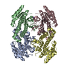
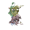
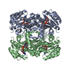
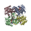
| Title | Crsytal structure of Acinetobacter baumannii FabG inhibitor complex at 2.35 A resolution | ||||||
 Components Components | 3-oxoacyl-(Acyl-carrier-protein) reductase | ||||||
 Keywords Keywords | BIOSYNTHETIC PROTEIN / Fatty acid biosynthesis / FabG / (3-oxoacyl-(Acyl-carrier-protein) reductase) / ligand complex / FAS-II | ||||||
| Function / homology |  Function and homology information Function and homology information3-oxoacyl-[acyl-carrier-protein] reductase / 3-oxoacyl-[acyl-carrier-protein] reductase (NADPH) activity / fatty acid elongation / NAD binding Similarity search - Function | ||||||
| Biological species |  Acinetobacter baumannii (bacteria) Acinetobacter baumannii (bacteria) | ||||||
| Method |  X-RAY DIFFRACTION / SYNCHROTRON / MOLECULAR REPLACEMENT / Resolution: 2.35 Å X-RAY DIFFRACTION / SYNCHROTRON / MOLECULAR REPLACEMENT / Resolution: 2.35 Å | ||||||
 Authors Authors | Rudraraju, R. / Schnell, R. / Schneider, G. | ||||||
| Funding support |  Sweden, 1items Sweden, 1items
| ||||||
 Citation Citation | Journal: Bioorg.Med.Chem. / Year: 2020 Title: A FabG inhibitor targeting an allosteric binding site inhibits several orthologs from Gram-negative ESKAPE pathogens. Authors: Vella, P. / Rudraraju, R.S. / Lundback, T. / Axelsson, H. / Almqvist, H. / Vallin, M. / Schneider, G. / Schnell, R. | ||||||
| History |
|
- Structure visualization
| Structure viewer | Molecule: MolmilJmol/JSmol |
|---|
- Downloads & links
-Download
| PDBx/mmCIF format | 6t65.cif.gz | 181.2 KB | Display | PDBx/mmCIF format |
|---|---|---|---|---|
| PDB format | pdb6t65.ent.gz | 143.6 KB | Display | PDB format |
| PDBx/mmJSON format | 6t65.json.gz | Tree view | PDBx/mmJSON format | |
| Others |  Other downloads Other downloads |
-Validation report
| Arichive directory | https://data.pdbj.org/pub/pdb/validation_reports/t6/6t65ftp://data.pdbj.org/pub/pdb/validation_reports/t6/6t65 | HTTPS FTP |
|---|
-Related structure data
| Related structure data |  6t5xC  6t60C  6t62C  6t6nC  6t6pC  6t77C  6t7mC  4afnS S: Starting model for refinement C: citing same article ( |
|---|---|
| Similar structure data |


-Links
 PDBj
PDBj


- Assembly
| Deposited unit | 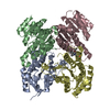
| ||||||||
|---|---|---|---|---|---|---|---|---|---|
| 1 |
| ||||||||
| Unit cell |
|
-Components
| #1: Protein | Mass: 26096.635 Da / Num. of mol.: 4 / Mutation: T2A Source method: isolated from a genetically manipulated source Details: The mutation T2A (position 2 in sequence, Thr mutated to Ala) was introduced for gene cloning reasons. Source: (gene. exp.) Acinetobacter baumannii (bacteria) / Gene: fabG / Production host: References: UniProt: V5VHN7, 3-oxoacyl-[acyl-carrier-protein] reductase #2: Chemical | ChemComp-MLH / |   Mass: 417.296 Da / Num. of mol.: 1 / Source method: obtained synthetically / Formula: C20H21BrN2O3 / Feature type: SUBJECT OF INVESTIGATION Mass: 417.296 Da / Num. of mol.: 1 / Source method: obtained synthetically / Formula: C20H21BrN2O3 / Feature type: SUBJECT OF INVESTIGATION#3: Water | ChemComp-HOH / |  Mass: 18.015 Da / Num. of mol.: 162 / Source method: isolated from a natural source / Formula: H2O Mass: 18.015 Da / Num. of mol.: 162 / Source method: isolated from a natural source / Formula: H2OHas ligand of interest | Y | |
|---|
-Experimental details
-Experiment
| Experiment | Method: X-RAY DIFFRACTION / Number of used crystals: 1 |
|---|
- Sample preparation
| Crystal | Density Matthews: 2.24 Å3/Da / Density % sol: 45.1 % |
|---|---|
| Crystal grow | Temperature: 293 K / Method: vapor diffusion, hanging drop / pH: 7.5 / Details: 0.2 M NaCl 0.1 M hepes pH 7.5 27% PEG 3350 |
-Data collection
| Diffraction | Mean temperature: 100 K / Serial crystal experiment: N |
|---|---|
| Diffraction source | Source: SYNCHROTRON / Site: ESRF  / Beamline: ID29 / Wavelength: 1.07227 Å / Beamline: ID29 / Wavelength: 1.07227 Å |
| Detector | Type: DECTRIS PILATUS 6M / Detector: PIXEL / Date: Apr 19, 2018 / Details: mirrors |
| Radiation | Monochromator: Si 111 / Protocol: SINGLE WAVELENGTH / Monochromatic (M) / Laue (L): M / Scattering type: x-ray |
| Radiation wavelength | Wavelength: 1.07227 Å / Relative weight: 1 |
| Reflection | Resolution: 2.35→44.08 Å / Num. obs: 38286 / % possible obs: 100 % / Observed criterion σ(I): 1.9 / Redundancy: 3.8 % / Biso Wilson estimate: 45.5 Å2 / CC1/2: 0.998 / Rmerge(I) obs: 0.053 / Rpim(I) all: 0.049 / Rrim(I) all: 0.071 / Rsym value: 0.053 / Net I/σ(I): 11.1 |
| Reflection shell | Resolution: 2.35→2.43 Å / Redundancy: 3.8 % / Rmerge(I) obs: 0.514 / Num. unique obs: 3749 / CC1/2: 0.81 / Rpim(I) all: 0.471 / Rrim(I) all: 0.7 / Rsym value: 0.598 / % possible all: 100 |
- Processing
| Software |
| ||||||||||||||||||||||||||||||||||||||||||||||||||||||||||||
|---|---|---|---|---|---|---|---|---|---|---|---|---|---|---|---|---|---|---|---|---|---|---|---|---|---|---|---|---|---|---|---|---|---|---|---|---|---|---|---|---|---|---|---|---|---|---|---|---|---|---|---|---|---|---|---|---|---|---|---|---|---|
| Refinement | Method to determine structure: MOLECULAR REPLACEMENT Starting model: 4AFN Resolution: 2.35→44 Å / Cor.coef. Fo:Fc: 0.952 / Cor.coef. Fo:Fc free: 0.946 / SU B: 8.631 / SU ML: 0.196 / Cross valid method: THROUGHOUT / σ(F): 0 / ESU R: 0.46 / ESU R Free: 0.251 Details: HYDROGENS HAVE BEEN ADDED IN THE RIDING POSITIONS U VALUES : REFINED INDIVIDUALLY
| ||||||||||||||||||||||||||||||||||||||||||||||||||||||||||||
| Solvent computation | Ion probe radii: 0.8 Å / Shrinkage radii: 0.8 Å / VDW probe radii: 1.2 Å | ||||||||||||||||||||||||||||||||||||||||||||||||||||||||||||
| Displacement parameters | Biso max: 146.33 Å2 / Biso mean: 48.348 Å2 / Biso min: 19.19 Å2
| ||||||||||||||||||||||||||||||||||||||||||||||||||||||||||||
| Refinement step | Cycle: final / Resolution: 2.35→44 Å
| ||||||||||||||||||||||||||||||||||||||||||||||||||||||||||||
| Refine LS restraints |
| ||||||||||||||||||||||||||||||||||||||||||||||||||||||||||||
| LS refinement shell | Resolution: 2.35→2.411 Å / Rfactor Rfree error: 0 / Total num. of bins used: 20
|
