 Movie
Movie Controller

[English] 日本語
 Yorodumi
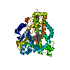
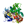
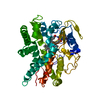
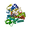
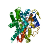
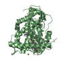
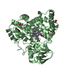
- PDB-6t0h: Crystal structure of CYP124 in complex with 1-alpha-hydroxy-vitamin D3 -
+ Open data

- Basic information
| Entry | Database: PDB / ID: 6t0h | |||||||||
|---|---|---|---|---|---|---|---|---|---|---|
| Title | Crystal structure of CYP124 in complex with 1-alpha-hydroxy-vitamin D3 | |||||||||
 Components Components | CYP124 in complex with inhibitor carbethoxyhexyl imidazole | |||||||||
 Keywords Keywords | OXIDOREDUCTASE / Cytochrome / P450 / CYP / 124 / CYP124 / vitamin / D3 / vitaminD3 / 1 alpha-hydroxy / 1-alpha-hydroxy / alfacalcidol / calciferol / cholecalciferol / tuberculosis / mycobacterium tuberculosis | |||||||||
| Function / homology |  Function and homology information Function and homology informationmethyl-branched lipid omega-hydroxylase / methyl-branched fatty acid metabolic process / cholesterol 26-hydroxylase activity / cholest-4-en-3-one 26-monooxygenase [(25R)-3-oxocholest-4-en-26-oate forming] / fatty acid omega-oxidation / cholest-4-en-3-one 26-monooxygenase activity / steroid hydroxylase activity / cholesterol catabolic process / NADPH binding / iron ion binding / heme binding Similarity search - Function | |||||||||
| Biological species |  Mycobacterium tuberculosis H37Rv (bacteria) Mycobacterium tuberculosis H37Rv (bacteria) | |||||||||
| Method |  X-RAY DIFFRACTION / SYNCHROTRON / MOLECULAR REPLACEMENT / Resolution: 1.18 Å X-RAY DIFFRACTION / SYNCHROTRON / MOLECULAR REPLACEMENT / Resolution: 1.18 Å | |||||||||
 Authors Authors | Bukhdruker, S. / Marin, E. / Varaksa, T. / Gilep, A. / Strushkevich, N. / Borshchevskiy, V. | |||||||||
| Funding support |  Russian Federation, 1items Russian Federation, 1items
| |||||||||
 Citation Citation | Journal: J.Mol.Biol. / Year: 2021 Title: Metabolic Fate of Human Immunoactive Sterols in Mycobacterium tuberculosis. Authors: Varaksa, T. / Bukhdruker, S. / Grabovec, I. / Marin, E. / Kavaleuski, A. / Gusach, A. / Kovalev, K. / Maslov, I. / Luginina, A. / Zabelskii, D. / Astashkin, R. / Shevtsov, M. / Smolskaya, S. ...Authors: Varaksa, T. / Bukhdruker, S. / Grabovec, I. / Marin, E. / Kavaleuski, A. / Gusach, A. / Kovalev, K. / Maslov, I. / Luginina, A. / Zabelskii, D. / Astashkin, R. / Shevtsov, M. / Smolskaya, S. / Kavaleuskaya, A. / Shabunya, P. / Baranovsky, A. / Dolgopalets, V. / Charnou, Y. / Savachka, A. / Litvinovskaya, R. / Hurski, A. / Shevchenko, E. / Rogachev, A. / Mishin, A. / Gordeliy, V. / Gabrielian, A. / Hurt, D.E. / Nikonenko, B. / Majorov, K. / Apt, A. / Rosenthal, A. / Gilep, A. / Borshchevskiy, V. / Strushkevich, N. | |||||||||
| History |
|
- Structure visualization
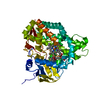
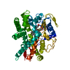
| Structure viewer | Molecule: MolmilJmol/JSmol |
|---|
- Downloads & links
-Download
| PDBx/mmCIF format | 6t0h.cif.gz | 289.9 KB | Display | PDBx/mmCIF format |
|---|---|---|---|---|
| PDB format | pdb6t0h.ent.gz | 205.9 KB | Display | PDB format |
| PDBx/mmJSON format | 6t0h.json.gz | Tree view | PDBx/mmJSON format | |
| Others |  Other downloads Other downloads |
-Validation report
| Arichive directory | https://data.pdbj.org/pub/pdb/validation_reports/t0/6t0hftp://data.pdbj.org/pub/pdb/validation_reports/t0/6t0h | HTTPS FTP |
|---|
-Related structure data
| Related structure data |  6t0fSC 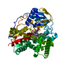 6t0gC 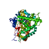 6t0kC  6t0lC S: Starting model for refinement C: citing same article ( |
|---|---|
| Similar structure data |
-Links
 PDBj
PDBj
- Assembly
| Deposited unit | 
| ||||||||||||
|---|---|---|---|---|---|---|---|---|---|---|---|---|---|
| 1 |
| ||||||||||||
| Unit cell |
|
-Components
-Protein , 1 types, 1 molecules A
| #1: Protein | Mass: 48838.930 Da / Num. of mol.: 1 Source method: isolated from a genetically manipulated source Source: (gene. exp.) Mycobacterium tuberculosis H37Rv (bacteria)Production host: References: UniProt: P9WPP3, methyl-branched lipid omega-hydroxylase, cholest-4-en-3-one 26-monooxygenase [(25R)-3-oxocholest-4-en-26-oate forming] |
|---|
-Non-polymers , 7 types, 742 molecules 


| #2: Chemical | ChemComp-HEM /  Mass: 616.487 Da / Num. of mol.: 1 / Source method: obtained synthetically / Formula: C34H32FeN4O4 Mass: 616.487 Da / Num. of mol.: 1 / Source method: obtained synthetically / Formula: C34H32FeN4O4 | ||||||||
|---|---|---|---|---|---|---|---|---|---|
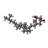
| #3: Chemical | ChemComp-M9B /  Mass: 400.637 Da / Num. of mol.: 1 / Source method: obtained synthetically / Formula: C27H44O2 / Feature type: SUBJECT OF INVESTIGATION / Comment: hormone*YM Mass: 400.637 Da / Num. of mol.: 1 / Source method: obtained synthetically / Formula: C27H44O2 / Feature type: SUBJECT OF INVESTIGATION / Comment: hormone*YM | ||||||||
| #4: Chemical | ChemComp-GOL /  Mass: 92.094 Da / Num. of mol.: 4 / Source method: obtained synthetically / Formula: C3H8O3 Mass: 92.094 Da / Num. of mol.: 4 / Source method: obtained synthetically / Formula: C3H8O3#5: Chemical | ChemComp-PGE /  Mass: 150.173 Da / Num. of mol.: 6 / Source method: obtained synthetically / Formula: C6H14O4 Mass: 150.173 Da / Num. of mol.: 6 / Source method: obtained synthetically / Formula: C6H14O4#6: Chemical |  Mass: 24.305 Da / Num. of mol.: 2 / Source method: obtained synthetically / Formula: Mg Mass: 24.305 Da / Num. of mol.: 2 / Source method: obtained synthetically / Formula: Mg#7: Chemical |  Mass: 35.453 Da / Num. of mol.: 3 / Source method: obtained synthetically / Formula: Cl Mass: 35.453 Da / Num. of mol.: 3 / Source method: obtained synthetically / Formula: Cl#8: Water | ChemComp-HOH / | Mass: 18.015 Da / Num. of mol.: 725 / Source method: isolated from a natural source / Formula: H2O |
-Details
| Has ligand of interest | Y |
|---|
-Experimental details
-Experiment
| Experiment | Method: X-RAY DIFFRACTION / Number of used crystals: 1 |
|---|
- Sample preparation
| Crystal | Density Matthews: 2.17 Å3/Da / Density % sol: 43.43 % |
|---|---|
| Crystal grow | Temperature: 293 K / Method: vapor diffusion, sitting drop / pH: 5.5 Details: 25% PEG 3350, 0.1M Bis-Tris, 0.2M Magnesium chloride |
-Data collection
| Diffraction | Mean temperature: 100 K / Serial crystal experiment: N | |||||||||||||||||||||||||||||||||||||||||||||||||||||||||||||||||||||||||||||||||||||||||||||||||||||||||||||||||||||||||||||||||||||||||||||||||||
|---|---|---|---|---|---|---|---|---|---|---|---|---|---|---|---|---|---|---|---|---|---|---|---|---|---|---|---|---|---|---|---|---|---|---|---|---|---|---|---|---|---|---|---|---|---|---|---|---|---|---|---|---|---|---|---|---|---|---|---|---|---|---|---|---|---|---|---|---|---|---|---|---|---|---|---|---|---|---|---|---|---|---|---|---|---|---|---|---|---|---|---|---|---|---|---|---|---|---|---|---|---|---|---|---|---|---|---|---|---|---|---|---|---|---|---|---|---|---|---|---|---|---|---|---|---|---|---|---|---|---|---|---|---|---|---|---|---|---|---|---|---|---|---|---|---|---|---|---|
| Diffraction source | Source: SYNCHROTRON / Site: ESRF  / Beamline: ID30B / Wavelength: 0.976 Å / Beamline: ID30B / Wavelength: 0.976 Å | |||||||||||||||||||||||||||||||||||||||||||||||||||||||||||||||||||||||||||||||||||||||||||||||||||||||||||||||||||||||||||||||||||||||||||||||||||
| Detector | Type: DECTRIS PILATUS3 6M / Detector: PIXEL / Date: Jun 6, 2018 | |||||||||||||||||||||||||||||||||||||||||||||||||||||||||||||||||||||||||||||||||||||||||||||||||||||||||||||||||||||||||||||||||||||||||||||||||||
| Radiation | Protocol: SINGLE WAVELENGTH / Monochromatic (M) / Laue (L): M / Scattering type: x-ray | |||||||||||||||||||||||||||||||||||||||||||||||||||||||||||||||||||||||||||||||||||||||||||||||||||||||||||||||||||||||||||||||||||||||||||||||||||
| Radiation wavelength | Wavelength: 0.976 Å / Relative weight: 1 | |||||||||||||||||||||||||||||||||||||||||||||||||||||||||||||||||||||||||||||||||||||||||||||||||||||||||||||||||||||||||||||||||||||||||||||||||||
| Reflection | Resolution: 1.05→20 Å / Num. obs: 186877 / % possible obs: 97.6 % / Redundancy: 3 % / Biso Wilson estimate: 10.87 Å2 / CC1/2: 0.993 / Rrim(I) all: 0.12 / Net I/σ(I): 5.7 | |||||||||||||||||||||||||||||||||||||||||||||||||||||||||||||||||||||||||||||||||||||||||||||||||||||||||||||||||||||||||||||||||||||||||||||||||||
| Reflection shell | Diffraction-ID: 1
|
- Processing
| Software |
| |||||||||||||||||||||||||||||||||||||||||||||||||||||||||||||||||||||||||||||||||||||||||||||||||||||||||||||||||||||||||||||||||||||||||||||||||||||||||||||||||||||||||||||||||||||||||||||||||||||||||||||||||||||||||
|---|---|---|---|---|---|---|---|---|---|---|---|---|---|---|---|---|---|---|---|---|---|---|---|---|---|---|---|---|---|---|---|---|---|---|---|---|---|---|---|---|---|---|---|---|---|---|---|---|---|---|---|---|---|---|---|---|---|---|---|---|---|---|---|---|---|---|---|---|---|---|---|---|---|---|---|---|---|---|---|---|---|---|---|---|---|---|---|---|---|---|---|---|---|---|---|---|---|---|---|---|---|---|---|---|---|---|---|---|---|---|---|---|---|---|---|---|---|---|---|---|---|---|---|---|---|---|---|---|---|---|---|---|---|---|---|---|---|---|---|---|---|---|---|---|---|---|---|---|---|---|---|---|---|---|---|---|---|---|---|---|---|---|---|---|---|---|---|---|---|---|---|---|---|---|---|---|---|---|---|---|---|---|---|---|---|---|---|---|---|---|---|---|---|---|---|---|---|---|---|---|---|---|---|---|---|---|---|---|---|---|---|---|---|---|---|---|---|---|
| Refinement | Method to determine structure: MOLECULAR REPLACEMENT Starting model: 6T0F Resolution: 1.18→19.72 Å / SU ML: 0.1406 / Cross valid method: FREE R-VALUE / σ(F): 1.34 / Phase error: 20.0532
| |||||||||||||||||||||||||||||||||||||||||||||||||||||||||||||||||||||||||||||||||||||||||||||||||||||||||||||||||||||||||||||||||||||||||||||||||||||||||||||||||||||||||||||||||||||||||||||||||||||||||||||||||||||||||
| Solvent computation | Shrinkage radii: 0.9 Å / VDW probe radii: 1.11 Å | |||||||||||||||||||||||||||||||||||||||||||||||||||||||||||||||||||||||||||||||||||||||||||||||||||||||||||||||||||||||||||||||||||||||||||||||||||||||||||||||||||||||||||||||||||||||||||||||||||||||||||||||||||||||||
| Displacement parameters | Biso mean: 18.17 Å2 | |||||||||||||||||||||||||||||||||||||||||||||||||||||||||||||||||||||||||||||||||||||||||||||||||||||||||||||||||||||||||||||||||||||||||||||||||||||||||||||||||||||||||||||||||||||||||||||||||||||||||||||||||||||||||
| Refinement step | Cycle: LAST / Resolution: 1.18→19.72 Å
| |||||||||||||||||||||||||||||||||||||||||||||||||||||||||||||||||||||||||||||||||||||||||||||||||||||||||||||||||||||||||||||||||||||||||||||||||||||||||||||||||||||||||||||||||||||||||||||||||||||||||||||||||||||||||
| Refine LS restraints |
| |||||||||||||||||||||||||||||||||||||||||||||||||||||||||||||||||||||||||||||||||||||||||||||||||||||||||||||||||||||||||||||||||||||||||||||||||||||||||||||||||||||||||||||||||||||||||||||||||||||||||||||||||||||||||
| LS refinement shell |
|
