 Movie
Movie Controller

[English] 日本語
 Yorodumi
- PDB-6rgg: Photorhabdus laumondii lectin PLL2 in complex with O-methylated P... -
+ Open data

- Basic information
| Entry | Database: PDB / ID: 6rgg | ||||||
|---|---|---|---|---|---|---|---|
| Title | Photorhabdus laumondii lectin PLL2 in complex with O-methylated PGL-1-derived disaccharide | ||||||
 Components Components | lectin PLL2 from Photorhabdus laumondii | ||||||
 Keywords Keywords | SUGAR BINDING PROTEIN / lectin / Photorhabdus / PGL-1 / O-methylated saccharide / beta-propeller | ||||||
| Function / homology | : / PLL-like beta propeller / Repeat of unknown function (DUF346) / 6-deoxy-2,3-di-O-methyl-alpha-L-mannopyranose / 3,6-O-dimethyl-D-glucose / Photorhabdus luminescens subsp. laumondii TTO1 complete genome segment 3/17 Function and homology information Function and homology information | ||||||
| Biological species |  Photorhabdus laumondii subsp. laumondii TTO1 (bacteria) Photorhabdus laumondii subsp. laumondii TTO1 (bacteria) | ||||||
| Method |  X-RAY DIFFRACTION / SYNCHROTRON / MOLECULAR REPLACEMENT / Resolution: 2.2 Å X-RAY DIFFRACTION / SYNCHROTRON / MOLECULAR REPLACEMENT / Resolution: 2.2 Å | ||||||
 Authors Authors | Houser, J. / Fujdiarova, E. / Wimmerova, M. | ||||||
 Citation Citation | Journal: Febs J. / Year: 2021 Title: Heptabladed beta-propeller lectins PLL2 and PHL from Photorhabdus spp. recognize O-methylated sugars and influence the host immune system. Authors: Fujdiarova, E. / Houser, J. / Dobes, P. / Paulikova, G. / Kondakov, N. / Kononov, L. / Hyrsl, P. / Wimmerova, M. | ||||||
| History |
|
- Structure visualization
| Structure viewer | Molecule: MolmilJmol/JSmol |
|---|
- Downloads & links
-Download
| PDBx/mmCIF format | 6rgg.cif.gz | 145.1 KB | Display | PDBx/mmCIF format |
|---|---|---|---|---|
| PDB format | pdb6rgg.ent.gz | 112.6 KB | Display | PDB format |
| PDBx/mmJSON format | 6rgg.json.gz | Tree view | PDBx/mmJSON format | |
| Others |  Other downloads Other downloads |
-Validation report
| Arichive directory | https://data.pdbj.org/pub/pdb/validation_reports/rg/6rggftp://data.pdbj.org/pub/pdb/validation_reports/rg/6rgg | HTTPS FTP |
|---|
-Related structure data
| Related structure data |  6rfzC  6rg1C  6rg2C 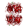 6rgjC  6rgrC  6rguC  6rgwC C: citing same article ( |
|---|---|
| Similar structure data |


-Links
 PDBj
PDBj- Assembly
| Deposited unit | 
| ||||||||
|---|---|---|---|---|---|---|---|---|---|
| 1 |
| ||||||||
| Unit cell |
|
-Components
-Protein / Sugars , 2 types, 4 molecules AB
| #1: Protein | Mass: 40150.957 Da / Num. of mol.: 2 Source method: isolated from a genetically manipulated source Source: (gene. exp.) Photorhabdus laumondii subsp. laumondii TTO1 (bacteria)Gene: plu0734 / Plasmid: pET25-b / Production host: #3: Sugar |  Type: L-saccharide, alpha linking / Mass: 192.210 Da / Num. of mol.: 2 / Source method: obtained synthetically / Formula: C8H16O5 Type: L-saccharide, alpha linking / Mass: 192.210 Da / Num. of mol.: 2 / Source method: obtained synthetically / Formula: C8H16O5 |
|---|
-Non-polymers , 4 types, 28 molecules 


| #2: Chemical | ChemComp-K3Q /  Mass: 208.209 Da / Num. of mol.: 6 / Source method: obtained synthetically / Formula: C8H16O6 Mass: 208.209 Da / Num. of mol.: 6 / Source method: obtained synthetically / Formula: C8H16O6#4: Chemical | ChemComp-NA /  Mass: 22.990 Da / Num. of mol.: 5 / Source method: obtained synthetically / Formula: Na Mass: 22.990 Da / Num. of mol.: 5 / Source method: obtained synthetically / Formula: Na#5: Chemical | ChemComp-EDO / |  Mass: 62.068 Da / Num. of mol.: 1 / Source method: obtained synthetically / Formula: C2H6O2 Mass: 62.068 Da / Num. of mol.: 1 / Source method: obtained synthetically / Formula: C2H6O2#6: Water | ChemComp-HOH / | Mass: 18.015 Da / Num. of mol.: 16 / Source method: isolated from a natural source / Formula: H2O |
|---|
-Experimental details
-Experiment
| Experiment | Method: X-RAY DIFFRACTION / Number of used crystals: 1 |
|---|
- Sample preparation
| Crystal | Density Matthews: 2.55 Å3/Da / Density % sol: 51.73 % |
|---|---|
| Crystal grow | Temperature: 293 K / Method: vapor diffusion, sitting drop / pH: 4.6 / Details: 12.5% PEG 4000, 0.1 M sodium acetate pH 4.6 |
-Data collection
| Diffraction | Mean temperature: 100 K / Serial crystal experiment: N |
|---|---|
| Diffraction source | Source: SYNCHROTRON / Site: PETRA III, EMBL c/o DESY  / Beamline: P13 (MX1) / Wavelength: 0.9763 Å / Beamline: P13 (MX1) / Wavelength: 0.9763 Å |
| Detector | Type: DECTRIS PILATUS 6M-F / Detector: PIXEL / Date: Oct 30, 2018 |
| Radiation | Protocol: SINGLE WAVELENGTH / Monochromatic (M) / Laue (L): M / Scattering type: x-ray |
| Radiation wavelength | Wavelength: 0.9763 Å / Relative weight: 1 |
| Reflection | Resolution: 2.2→67.03 Å / Num. obs: 40910 / % possible obs: 99.8 % / Redundancy: 3.8 % / Biso Wilson estimate: 46.387 Å2 / CC1/2: 0.993 / Rmerge(I) obs: 0.096 / Net I/σ(I): 5.1 |
| Reflection shell | Resolution: 2.2→2.32 Å / Redundancy: 3.8 % / Rmerge(I) obs: 0.922 / Mean I/σ(I) obs: 1.1 / Num. unique obs: 5878 / CC1/2: 0.541 / % possible all: 99.8 |
- Processing
| Software |
| ||||||||||||||||||||||||||||||||||||||||||||||||||||||||||||||||||||||||||||||||||||||||||||||||||||||||||||||||||||||||||||||||||||||||||||||||||||||||||||||||||||||||||||||||||||||
|---|---|---|---|---|---|---|---|---|---|---|---|---|---|---|---|---|---|---|---|---|---|---|---|---|---|---|---|---|---|---|---|---|---|---|---|---|---|---|---|---|---|---|---|---|---|---|---|---|---|---|---|---|---|---|---|---|---|---|---|---|---|---|---|---|---|---|---|---|---|---|---|---|---|---|---|---|---|---|---|---|---|---|---|---|---|---|---|---|---|---|---|---|---|---|---|---|---|---|---|---|---|---|---|---|---|---|---|---|---|---|---|---|---|---|---|---|---|---|---|---|---|---|---|---|---|---|---|---|---|---|---|---|---|---|---|---|---|---|---|---|---|---|---|---|---|---|---|---|---|---|---|---|---|---|---|---|---|---|---|---|---|---|---|---|---|---|---|---|---|---|---|---|---|---|---|---|---|---|---|---|---|---|---|
| Refinement | Method to determine structure: MOLECULAR REPLACEMENT / Resolution: 2.2→67.03 Å / Cor.coef. Fo:Fc: 0.959 / Cor.coef. Fo:Fc free: 0.945 / Cross valid method: THROUGHOUT / ESU R: 0.249 / ESU R Free: 0.187 / Details: HYDROGENS HAVE BEEN ADDED IN THE RIDING POSITIONS
| ||||||||||||||||||||||||||||||||||||||||||||||||||||||||||||||||||||||||||||||||||||||||||||||||||||||||||||||||||||||||||||||||||||||||||||||||||||||||||||||||||||||||||||||||||||||
| Solvent computation | Ion probe radii: 0.8 Å / Shrinkage radii: 0.8 Å / VDW probe radii: 1.2 Å | ||||||||||||||||||||||||||||||||||||||||||||||||||||||||||||||||||||||||||||||||||||||||||||||||||||||||||||||||||||||||||||||||||||||||||||||||||||||||||||||||||||||||||||||||||||||
| Displacement parameters | Biso mean: 62.054 Å2
| ||||||||||||||||||||||||||||||||||||||||||||||||||||||||||||||||||||||||||||||||||||||||||||||||||||||||||||||||||||||||||||||||||||||||||||||||||||||||||||||||||||||||||||||||||||||
| Refinement step | Cycle: 1 / Resolution: 2.2→67.03 Å
| ||||||||||||||||||||||||||||||||||||||||||||||||||||||||||||||||||||||||||||||||||||||||||||||||||||||||||||||||||||||||||||||||||||||||||||||||||||||||||||||||||||||||||||||||||||||
| Refine LS restraints |
|