 Movie
Movie Controller

[English] 日本語
 Yorodumi
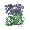
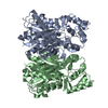
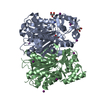
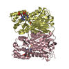
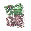
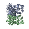
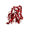
- PDB-6j1n: Anisodus acutangulus type III polyketide sythase AaPKS2 in comple... -
+ Open data

- Basic information
| Entry | Database: PDB / ID: 6j1n | ||||||
|---|---|---|---|---|---|---|---|
| Title | Anisodus acutangulus type III polyketide sythase AaPKS2 in complex with 4-carboxy-3-oxobutanoyl-CoA | ||||||
 Components Components | A. acutangulus PKS2 | ||||||
 Keywords Keywords | BIOSYNTHETIC PROTEIN / Anisodus acutangulus / type III polyketide sythase / tropane alkaloids biosynthesis | ||||||
| Function / homology | Thiolase/Chalcone synthase / Peroxisomal Thiolase; Chain A, domain 1 / 3-Layer(aba) Sandwich / Alpha Beta / Chem-B7X Function and homology information Function and homology information | ||||||
| Biological species |  Anisodus acutangulus (plant) Anisodus acutangulus (plant) | ||||||
| Method |  X-RAY DIFFRACTION / SYNCHROTRON / MOLECULAR REPLACEMENT / Resolution: 2.532 Å X-RAY DIFFRACTION / SYNCHROTRON / MOLECULAR REPLACEMENT / Resolution: 2.532 Å | ||||||
 Authors Authors | Fang, C.L. / Zhang, Y. | ||||||
| Funding support |  China, 1items China, 1items
| ||||||
 Citation Citation | Journal: Nat Commun / Year: 2019 Title: Tropane alkaloids biosynthesis involves an unusual type III polyketide synthase and non-enzymatic condensation. Authors: Huang, J.P. / Fang, C. / Ma, X. / Wang, L. / Yang, J. / Luo, J. / Yan, Y. / Zhang, Y. / Huang, S.X. | ||||||
| History |
|
- Structure visualization
| Structure viewer | Molecule: MolmilJmol/JSmol |
|---|
- Downloads & links
-Download
| PDBx/mmCIF format | 6j1n.cif.gz | 170 KB | Display | PDBx/mmCIF format |
|---|---|---|---|---|
| PDB format | pdb6j1n.ent.gz | 130.9 KB | Display | PDB format |
| PDBx/mmJSON format | 6j1n.json.gz | Tree view | PDBx/mmJSON format | |
| Others |  Other downloads Other downloads |
-Validation report
| Arichive directory | https://data.pdbj.org/pub/pdb/validation_reports/j1/6j1nftp://data.pdbj.org/pub/pdb/validation_reports/j1/6j1n | HTTPS FTP |
|---|
-Related structure data
| Related structure data | 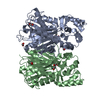 6j1mC  1cmlS S: Starting model for refinement C: citing same article ( |
|---|---|
| Similar structure data |


-Links
 PDBj
PDBj- Assembly
| Deposited unit | 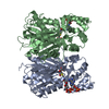
| ||||||||
|---|---|---|---|---|---|---|---|---|---|
| 1 |
| ||||||||
| Unit cell |
|
-Components
| #1: Protein | Mass: 46726.852 Da / Num. of mol.: 2 Source method: isolated from a genetically manipulated source Source: (gene. exp.) Anisodus acutangulus (plant) / Production host:  #2: Chemical | 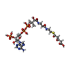  Mass: 895.617 Da / Num. of mol.: 2 / Source method: obtained synthetically / Formula: C26H40N7O20P3S Mass: 895.617 Da / Num. of mol.: 2 / Source method: obtained synthetically / Formula: C26H40N7O20P3S#3: Water | ChemComp-HOH / |  Mass: 18.015 Da / Num. of mol.: 404 / Source method: isolated from a natural source / Formula: H2O Mass: 18.015 Da / Num. of mol.: 404 / Source method: isolated from a natural source / Formula: H2OHas protein modification | Y | |
|---|
-Experimental details
-Experiment
| Experiment | Method: X-RAY DIFFRACTION / Number of used crystals: 1 |
|---|
- Sample preparation
| Crystal | Density Matthews: 3.47 Å3/Da / Density % sol: 64.57 % |
|---|---|
| Crystal grow | Temperature: 277.15 K / Method: vapor diffusion, sitting drop Details: 1% tryptone, 0.05M HEPES 7.0, 20% PEG3350, 0.001M Sodium azide |
-Data collection
| Diffraction | Mean temperature: 100 K / Serial crystal experiment: N |
|---|---|
| Diffraction source | Source: SYNCHROTRON / Site: SSRF / Beamline: BL17U1 / Wavelength: 0.979 Å |
| Detector | Type: DECTRIS EIGER X 16M / Detector: PIXEL / Date: Oct 28, 2018 |
| Radiation | Protocol: SINGLE WAVELENGTH / Monochromatic (M) / Laue (L): M / Scattering type: x-ray |
| Radiation wavelength | Wavelength: 0.979 Å / Relative weight: 1 |
| Reflection | Resolution: 2.53→50 Å / Num. obs: 44048 / % possible obs: 100 % / Observed criterion σ(F): 0 / Redundancy: 16.4 % / CC1/2: 0.998 / Rmerge(I) obs: 0.092 / Rpim(I) all: 0.024 / Rrim(I) all: 0.095 / Rsym value: 0.092 / Χ2: 0.96 / Net I/σ(I): 34.1 |
| Reflection shell | Resolution: 2.53→2.57 Å / Redundancy: 16.4 % / Rmerge(I) obs: 0.472 / Num. unique obs: 2176 / CC1/2: 0.974 / Rpim(I) all: 0.122 / Rrim(I) all: 0.488 / Rsym value: 0.472 / Χ2: 0.921 / % possible all: 100 |
- Processing
| Software |
| ||||||||||||||||||||||||||||||||||||||||||||||||||||||||||||||||||||||||||||||||||||||||||
|---|---|---|---|---|---|---|---|---|---|---|---|---|---|---|---|---|---|---|---|---|---|---|---|---|---|---|---|---|---|---|---|---|---|---|---|---|---|---|---|---|---|---|---|---|---|---|---|---|---|---|---|---|---|---|---|---|---|---|---|---|---|---|---|---|---|---|---|---|---|---|---|---|---|---|---|---|---|---|---|---|---|---|---|---|---|---|---|---|---|---|---|
| Refinement | Method to determine structure: MOLECULAR REPLACEMENT Starting model: 1CML Resolution: 2.532→47.149 Å / SU ML: 0.23 / Cross valid method: THROUGHOUT / σ(F): 1.52 / Phase error: 18.02 / Stereochemistry target values: ML
| ||||||||||||||||||||||||||||||||||||||||||||||||||||||||||||||||||||||||||||||||||||||||||
| Solvent computation | Shrinkage radii: 0.9 Å / VDW probe radii: 1.11 Å / Solvent model: FLAT BULK SOLVENT MODEL | ||||||||||||||||||||||||||||||||||||||||||||||||||||||||||||||||||||||||||||||||||||||||||
| Displacement parameters | Biso max: 93.75 Å2 / Biso mean: 33.6874 Å2 / Biso min: 15.13 Å2 | ||||||||||||||||||||||||||||||||||||||||||||||||||||||||||||||||||||||||||||||||||||||||||
| Refinement step | Cycle: final / Resolution: 2.532→47.149 Å
| ||||||||||||||||||||||||||||||||||||||||||||||||||||||||||||||||||||||||||||||||||||||||||
| LS refinement shell | Refine-ID: X-RAY DIFFRACTION / Rfactor Rfree error: 0 / Total num. of bins used: 14 / % reflection obs: 100 %
|
