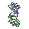
 Movie
Movie Controller


+ Open data

- Basic information
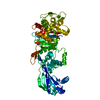
| Entry | Database: PDB / ID: 6g9s | ||||||
|---|---|---|---|---|---|---|---|
| Title | Structural basis for the inhibition of E. coli PBP2 | ||||||
 Components Components | Peptidoglycan D,D-transpeptidase MrdA | ||||||
 Keywords Keywords | HYDROLASE/ANTIBIOTIC / penicillin binding protein / HYDROLASE-ANTIBIOTIC complex | ||||||
| Function / homology |  Function and homology information Function and homology informationpeptidoglycan L,D-transpeptidase activity / serine-type D-Ala-D-Ala carboxypeptidase / serine-type D-Ala-D-Ala carboxypeptidase activity / penicillin binding / peptidoglycan biosynthetic process / cell wall organization / regulation of cell shape / outer membrane-bounded periplasmic space / response to antibiotic / proteolysis / plasma membrane Similarity search - Function | ||||||
| Biological species |  | ||||||
| Method |  X-RAY DIFFRACTION / SYNCHROTRON / MOLECULAR REPLACEMENT / Resolution: 2.001 Å X-RAY DIFFRACTION / SYNCHROTRON / MOLECULAR REPLACEMENT / Resolution: 2.001 Å | ||||||
 Authors Authors | Ruff, M. / Levy, N. | ||||||
 Citation Citation | Journal: J.Med.Chem. / Year: 2019 Title: Structural Basis for E. coli Penicillin Binding Protein (PBP) 2 Inhibition, a Platform for Drug Design. Authors: Levy, N. / Bruneau, J.M. / Le Rouzic, E. / Bonnard, D. / Le Strat, F. / Caravano, A. / Chevreuil, F. / Barbion, J. / Chasset, S. / Ledoussal, B. / Moreau, F. / Ruff, M. | ||||||
| History |
|
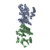
- Structure visualization
| Structure viewer | Molecule: MolmilJmol/JSmol |
|---|
- Downloads & links
-Download
| PDBx/mmCIF format | 6g9s.cif.gz | 235.9 KB | Display | PDBx/mmCIF format |
|---|---|---|---|---|
| PDB format | pdb6g9s.ent.gz | 187.9 KB | Display | PDB format |
| PDBx/mmJSON format | 6g9s.json.gz | Tree view | PDBx/mmJSON format | |
| Others |  Other downloads Other downloads |
-Validation report
| Arichive directory | https://data.pdbj.org/pub/pdb/validation_reports/g9/6g9sftp://data.pdbj.org/pub/pdb/validation_reports/g9/6g9s | HTTPS FTP |
|---|
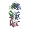
-Related structure data
| Related structure data | 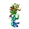 6g9fC  6g9pC  1vqqS  1xkwS 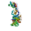 3oclS  4mnrS  4oolS  4wejS 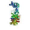 4wekS 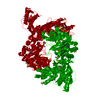 5dvyS 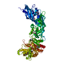 5e31S C: citing same article ( S: Starting model for refinement |
|---|---|
| Similar structure data |


-Links
 PDBj
PDBj
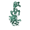
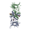
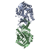
- Assembly
| Deposited unit | 
| ||||||||
|---|---|---|---|---|---|---|---|---|---|
| 1 |
| ||||||||
| Unit cell |
| ||||||||
| Components on special symmetry positions |
|
-Components
| #1: Protein | Mass: 65075.520 Da / Num. of mol.: 1 Source method: isolated from a genetically manipulated source Details: Serine 330 covalently modified with ligand / Source: (gene. exp.) References: UniProt: P0AD65, serine-type D-Ala-D-Ala carboxypeptidase |
|---|---|
| #2: Chemical | ChemComp-ET5 / (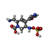  Mass: 334.306 Da / Num. of mol.: 1 / Source method: obtained synthetically / Formula: C10H14N4O7S Mass: 334.306 Da / Num. of mol.: 1 / Source method: obtained synthetically / Formula: C10H14N4O7S |
| #3: Water | ChemComp-HOH /  Mass: 18.015 Da / Num. of mol.: 364 / Source method: isolated from a natural source / Formula: H2O Mass: 18.015 Da / Num. of mol.: 364 / Source method: isolated from a natural source / Formula: H2O |
| Has protein modification | Y |
-Experimental details
-Experiment
| Experiment | Method: X-RAY DIFFRACTION / Number of used crystals: 1 |
|---|
- Sample preparation
| Crystal | Density Matthews: 3.6 Å3/Da / Density % sol: 65.8 % |
|---|---|
| Crystal grow | Temperature: 293 K / Method: vapor diffusion, hanging drop / pH: 8.5 Details: 0.1 M TrisBase / Bicine pH 8,5 ; 0.1 M Carboxylic acids mix (0.02 M Sodium formate ; 0.02 M Ammonium acetate ; 0.02 M Sodium citrate tribasic dihydrate ; 0.02 M Sodium potassium tartrate ...Details: 0.1 M TrisBase / Bicine pH 8,5 ; 0.1 M Carboxylic acids mix (0.02 M Sodium formate ; 0.02 M Ammonium acetate ; 0.02 M Sodium citrate tribasic dihydrate ; 0.02 M Sodium potassium tartrate tetrahydrate ; 0.02 M Sodium oxamate) ; 24 % Glycerol ; 12 % PEG 4000 |
-Data collection
| Diffraction | Mean temperature: 100 K |
|---|---|
| Diffraction source | Source: SYNCHROTRON / Site: SLS  / Beamline: X06DA / Wavelength: 1 Å / Beamline: X06DA / Wavelength: 1 Å |
| Detector | Type: DECTRIS PILATUS 2M / Detector: PIXEL / Date: Nov 29, 2016 |
| Radiation | Protocol: SINGLE WAVELENGTH / Monochromatic (M) / Laue (L): M / Scattering type: x-ray |
| Radiation wavelength | Wavelength: 1 Å / Relative weight: 1 |
| Reflection | Resolution: 2→45.7 Å / Num. obs: 59191 / % possible obs: 92.84 % / Redundancy: 11.3 % / CC1/2: 0.995 / Rpim(I) all: 0.05733 / Rrim(I) all: 0.1985 / Net I/σ(I): 8.32 |
| Reflection shell | Resolution: 2→2.07 Å / Redundancy: 10.4 % / Rmerge(I) obs: 3.254 / Num. unique obs: 4729 / CC1/2: 0.57 / Rpim(I) all: 1.054 / Rrim(I) all: 3.424 / % possible all: 80.6 |
- Processing
| Software |
| ||||||||||||||||||||||||||||||||||||||||||||||||||||||||||||||||||||||||||||||||||||||||||||||||||
|---|---|---|---|---|---|---|---|---|---|---|---|---|---|---|---|---|---|---|---|---|---|---|---|---|---|---|---|---|---|---|---|---|---|---|---|---|---|---|---|---|---|---|---|---|---|---|---|---|---|---|---|---|---|---|---|---|---|---|---|---|---|---|---|---|---|---|---|---|---|---|---|---|---|---|---|---|---|---|---|---|---|---|---|---|---|---|---|---|---|---|---|---|---|---|---|---|---|---|---|
| Refinement | Method to determine structure: MOLECULAR REPLACEMENT Starting model: ensemble of models was used: 5e31, 5dvy, 1vqq, 3ocl, 4ool, 4wek, 4wej, 4mnr, 1xkw Resolution: 2.001→44.405 Å / SU ML: 0.41 / Cross valid method: FREE R-VALUE / σ(F): 0 / Phase error: 33.03
| ||||||||||||||||||||||||||||||||||||||||||||||||||||||||||||||||||||||||||||||||||||||||||||||||||
| Solvent computation | Shrinkage radii: 0.9 Å / VDW probe radii: 1.11 Å | ||||||||||||||||||||||||||||||||||||||||||||||||||||||||||||||||||||||||||||||||||||||||||||||||||
| Refinement step | Cycle: LAST / Resolution: 2.001→44.405 Å
| ||||||||||||||||||||||||||||||||||||||||||||||||||||||||||||||||||||||||||||||||||||||||||||||||||
| Refine LS restraints |
| ||||||||||||||||||||||||||||||||||||||||||||||||||||||||||||||||||||||||||||||||||||||||||||||||||
| LS refinement shell |
| ||||||||||||||||||||||||||||||||||||||||||||||||||||||||||||||||||||||||||||||||||||||||||||||||||
| Refinement TLS params. | Method: refined / Origin x: 29.2097 Å / Origin y: 55.0645 Å / Origin z: 38.9349 Å
| ||||||||||||||||||||||||||||||||||||||||||||||||||||||||||||||||||||||||||||||||||||||||||||||||||
| Refinement TLS group | Selection details: all |
