 Movie
Movie Controller


+ Open data

- Basic information
| Entry |  Database: PDB chemical components / ID: 6AP Database: PDB chemical components / ID: 6AP |
|---|---|
| Name | Name: |
-Chemical information
| Composition |  | ||||||
|---|---|---|---|---|---|---|---|
| Others | Type: NON-POLYMER / PDB classification: HETAIN / Three letter code: 6AP / Model coordinates PDB-ID: 2B57 | ||||||
| History |
| ||||||
 External links External links | UniChem / ChemSpider / BindingDB / Brenda / ChEBI / ChEMBL / CompTox / HMDB / PubChem / SureChEMBL / Wikipedia search / Google search |
- Structure visualization
| Structure viewer | Molecule:  MolmilJmol/JSmol MolmilJmol/JSmol |
|---|
-Details
-SMILES
| ACDLabs 10.04 | | CACTVS 3.341 | OpenEye OEToolkits 1.5.0 | |
|---|
-SMILES CANONICAL
| CACTVS 3.341 | | OpenEye OEToolkits 1.5.0 | |
|---|
-InChI
| InChI 1.03 |
|---|
-InChIKey
| InChI 1.03 |
|---|
-SYSTEMATIC NAME
| ACDLabs 10.04 | | OpenEye OEToolkits 1.5.0 | |
|---|
-PDB entries
Showing all 5 items
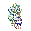
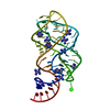
PDB-2b57: 
Guanine Riboswitch C74U mutant bound to 2,6-diaminopurine

PDB-4feo:
Crystal structure of the AU25A/A46G/C74U mutant xpt-pbuX guanine riboswitch aptamer domain in complex with 2,6-diaminopurine
PDB-4fep:
Crystal structure of the A24U/U25A/A46G/C74U mutant xpt-pbuX guanine riboswitch aptamer domain in complex with 2,6-diaminopurine

PDB-4lcp:
Crytsal structure of NE0047 in complex with 2,6-diaminopurine

PDB-4lvz:
Structure of the THF riboswitch bound to 2,6-diaminopurine
