 Movie
Movie Controller

[English] 日本語
 Yorodumi
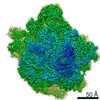
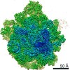
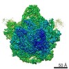
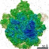
- EMDB-6711: Structure of the 50S large subunit of chloroplast ribosome from s... -
+ Open data

- Basic information
| Entry | Database: EMDB / ID: EMD-6711 | |||||||||
|---|---|---|---|---|---|---|---|---|---|---|
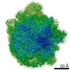
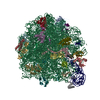
| Title | Structure of the 50S large subunit of chloroplast ribosome from spinach | |||||||||
 Map data Map data | ||||||||||
 Sample Sample |
| |||||||||
 Keywords Keywords | Cryo-EM / ribosome / chloroplast ribosome | |||||||||
| Function / homology |  Function and homology information Function and homology informationplastid translation / chloroplast envelope / mitochondrial large ribosomal subunit / mitochondrial translation / chloroplast thylakoid membrane / chloroplast / DNA-templated transcription termination / large ribosomal subunit / transferase activity / 5S rRNA binding ...plastid translation / chloroplast envelope / mitochondrial large ribosomal subunit / mitochondrial translation / chloroplast thylakoid membrane / chloroplast / DNA-templated transcription termination / large ribosomal subunit / transferase activity / 5S rRNA binding / ribosomal large subunit assembly / large ribosomal subunit rRNA binding / cytosolic large ribosomal subunit / negative regulation of translation / rRNA binding / structural constituent of ribosome / ribosome / translation / ribonucleoprotein complex / mRNA binding / mitochondrion / RNA binding Similarity search - Function | |||||||||
| Biological species |  Spinacia oleracea (spinach) Spinacia oleracea (spinach) | |||||||||
| Method | single particle reconstruction / cryo EM / Resolution: 3.3 Å | |||||||||
 Authors Authors | Ahmed T / Shi J | |||||||||
| Funding support |  Singapore, 1 items Singapore, 1 items
| |||||||||
 Citation Citation | Journal: Nucleic Acids Res / Year: 2017 Title: Unique localization of the plastid-specific ribosomal proteins in the chloroplast ribosome small subunit provides mechanistic insights into the chloroplastic translation. Authors: Tofayel Ahmed / Jian Shi / Shashi Bhushan / Abstract: Chloroplastic translation is mediated by a bacterial-type 70S chloroplast ribosome. During the evolution, chloroplast ribosomes have acquired five plastid-specific ribosomal proteins or PSRPs (cS22, ...Chloroplastic translation is mediated by a bacterial-type 70S chloroplast ribosome. During the evolution, chloroplast ribosomes have acquired five plastid-specific ribosomal proteins or PSRPs (cS22, cS23, bTHXc, cL37 and cL38) which have been suggested to play important regulatory roles in translation. However, their exact locations on the chloroplast ribosome remain elusive due to lack of a high-resolution structure, hindering our progress to understand their possible roles. Here we present a cryo-EM structure of the 70S chloroplast ribosome from spinach resolved to 3.4 Å and focus our discussion mainly on the architecture of the 30S small subunit (SSU) which is resolved to 3.7 Å. cS22 localizes at the SSU foot where it seems to compensate for the deletions in 16S rRNA. The mRNA exit site is highly remodeled due to the presence of cS23 suggesting an alternative mode of translation initiation. bTHXc is positioned at the SSU head and appears to stabilize the intersubunit bridge B1b during thermal fluctuations. The translation factor plastid pY binds to the SSU on the intersubunit side and interacts with the conserved nucleotide bases involved in decoding. Most of the intersubunit bridges are conserved compared to the bacteria, except for a new bridge involving uL2c and bS6c. | |||||||||
| History |
|
- Structure visualization
| Movie |
Movie viewer |
|---|---|
| Structure viewer | EM map: SurfViewMolmilJmol/JSmol |
| Supplemental images |

- Downloads & links
-EMDB archive
| Map data | emd_6711.map.gz | 22.5 MB | EMDB map data format | |
|---|---|---|---|---|
| Header (meta data) | emd-6711-v30.xmlemd-6711.xml | 44.8 KB 44.8 KB | Display Display | EMDB header |
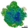
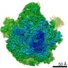
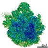
| Images | 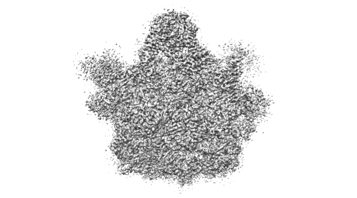 emd_6711.png emd_6711.png | 133.7 KB | ||
| Filedesc metadata | emd-6711.cif.gz | 10.4 KB | ||
| Archive directory |  http://ftp.pdbj.org/pub/emdb/structures/EMD-6711ftp://ftp.pdbj.org/pub/emdb/structures/EMD-6711 http://ftp.pdbj.org/pub/emdb/structures/EMD-6711ftp://ftp.pdbj.org/pub/emdb/structures/EMD-6711 | HTTPS FTP |
-Related structure data
| Related structure data |  5x8tMC 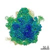 6709C 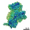 6710C 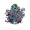 5x8pC 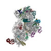 5x8rC M: atomic model generated by this map C: citing same article ( |
|---|---|
| Similar structure data |

-Links
| EMDB pages | EMDB (EBI/PDBe) / EMDataResource |
|---|---|
| Related items in Molecule of the Month |


-Map
| File | Download / File: emd_6711.map.gz / Format: CCP4 / Size: 216 MB / Type: IMAGE STORED AS FLOATING POINT NUMBER (4 BYTES) | ||||||||||||||||||||||||||||||||||||||||||||||||||||||||||||
|---|---|---|---|---|---|---|---|---|---|---|---|---|---|---|---|---|---|---|---|---|---|---|---|---|---|---|---|---|---|---|---|---|---|---|---|---|---|---|---|---|---|---|---|---|---|---|---|---|---|---|---|---|---|---|---|---|---|---|---|---|---|
| Projections & slices | Image control
Images are generated by Spider. | ||||||||||||||||||||||||||||||||||||||||||||||||||||||||||||
| Voxel size | X=Y=Z: 1.05 Å | ||||||||||||||||||||||||||||||||||||||||||||||||||||||||||||
| Density |
| ||||||||||||||||||||||||||||||||||||||||||||||||||||||||||||
| Symmetry | Space group: 1 | ||||||||||||||||||||||||||||||||||||||||||||||||||||||||||||
| Details | EMDB XML:
CCP4 map header:
| ||||||||||||||||||||||||||||||||||||||||||||||||||||||||||||
 Z (Sec.)
 Y (Row.)
 X (Col.)


-Supplemental data
- Sample components
+Entire : 50S large subunit of chloroplast ribosome from spinach
+Supramolecule #1: 50S large subunit of chloroplast ribosome from spinach
+Macromolecule #1: 50S ribosomal protein L32, chloroplastic
+Macromolecule #2: 50S ribosomal protein L33, chloroplastic
+Macromolecule #3: 50S ribosomal protein L34, chloroplastic
+Macromolecule #4: 50S ribosomal protein L35, chloroplastic
+Macromolecule #5: 50S ribosomal protein L36, chloroplastic
+Macromolecule #6: protein cL37
+Macromolecule #7: protein cL38
+Macromolecule #9: 50S ribosomal protein L2, chloroplastic
+Macromolecule #10: protein L3
+Macromolecule #11: 50S ribosomal protein L4, chloroplastic
+Macromolecule #12: 50S ribosomal protein L5, chloroplastic
+Macromolecule #13: protein L6
+Macromolecule #14: protein L9
+Macromolecule #15: 50S ribosomal protein L13, chloroplastic
+Macromolecule #16: 50S ribosomal protein L14, chloroplastic
+Macromolecule #17: protein L15
+Macromolecule #18: 50S ribosomal protein L16, chloroplastic
+Macromolecule #19: protein L17
+Macromolecule #20: protein L18
+Macromolecule #21: 50S ribosomal protein L19, chloroplastic
+Macromolecule #22: 50S ribosomal protein L20, chloroplastic
+Macromolecule #23: 50S ribosomal protein L21, chloroplastic
+Macromolecule #24: 50S ribosomal protein L22, chloroplastic
+Macromolecule #25: 50S ribosomal protein L23, chloroplastic
+Macromolecule #26: 50S ribosomal protein L24, chloroplastic
+Macromolecule #28: protein L27
+Macromolecule #29: protein L28
+Macromolecule #30: protein L29
+Macromolecule #32: 50S ribosomal protein L31
+Macromolecule #8: 5S rRNA
+Macromolecule #27: 4.8S rRNA
+Macromolecule #31: 23S rRNA
-Experimental details
-Structure determination
| Method | cryo EM |
|---|---|
 Processing Processing | single particle reconstruction |
| Aggregation state | particle |
-Sample preparation
| Buffer | pH: 7.6 Details: 20 mM Tris HCl, pH 7.6, 100 mM KCl, 10 mM MgOAc, 100 mM sucrose, 7 mM 2-mercaptoethanol, 1 unit/ml RNase inhibitor, 0.1% protease inhibitor |
|---|---|
| Grid | Model: Quantifoil / Material: COPPER / Mesh: 300 / Support film - Material: CARBON / Support film - topology: CONTINUOUS / Support film - Film thickness: 2 / Pretreatment - Type: GLOW DISCHARGE / Pretreatment - Time: 30 sec. |
| Vitrification | Cryogen name: ETHANE / Chamber humidity: 100 % / Chamber temperature: 277 K / Instrument: FEI VITROBOT MARK IV |
- Electron microscopy
| Microscope | FEI TITAN KRIOS |
|---|---|
| Image recording | Film or detector model: FEI FALCON II (4k x 4k) / Detector mode: INTEGRATING / Digitization - Dimensions - Width: 4096 pixel / Digitization - Dimensions - Height: 4096 pixel / Digitization - Frames/image: 1-25 / Number grids imaged: 1 / Number real images: 3161 / Average electron dose: 1.5 e/Å2 |
| Electron beam | Acceleration voltage: 300 kV / Electron source:  FIELD EMISSION GUN FIELD EMISSION GUN |
| Electron optics | C2 aperture diameter: 100.0 µm / Calibrated defocus max: 3.7 µm / Calibrated defocus min: 0.4 µm / Calibrated magnification: 133333 / Illumination mode: SPOT SCAN / Imaging mode: BRIGHT FIELD / Cs: 2.7 mm / Nominal defocus max: 3.7 µm / Nominal defocus min: 0.4 µm / Nominal magnification: 75000 |
| Sample stage | Specimen holder model: FEI TITAN KRIOS AUTOGRID HOLDER / Cooling holder cryogen: NITROGEN |
| Experimental equipment |  Model: Titan Krios / Image courtesy: FEI Company |
