 Movie
Movie Controller

[English] 日本語
 Yorodumi
- PDB-5zvt: Structure of RNA polymerase complex and genome within a dsRNA vir... -
+ Open data

- Basic information
| Entry | Database: PDB / ID: 5zvt | ||||||||||||||||||||||||||||||||||||
|---|---|---|---|---|---|---|---|---|---|---|---|---|---|---|---|---|---|---|---|---|---|---|---|---|---|---|---|---|---|---|---|---|---|---|---|---|---|
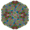
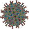
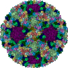
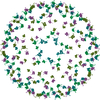
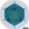
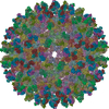
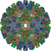
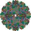
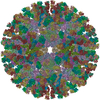
| Title | Structure of RNA polymerase complex and genome within a dsRNA virus provides insights into the mechanisms of transcription and assembly | ||||||||||||||||||||||||||||||||||||
 Components Components |
| ||||||||||||||||||||||||||||||||||||
 Keywords Keywords | VIRUS / icosahedral capsid / symmetry-mismatch / genome / RNA-dependent RNA polymerase | ||||||||||||||||||||||||||||||||||||
| Function / homology |  Function and homology information Function and homology informationsymbiont entry into host cell via permeabilization of inner membrane / permeabilization of host organelle membrane involved in viral entry into host cell / host cell surface binding / viral inner capsid / viral outer capsid / 7-methylguanosine mRNA capping / viral capsid / mRNA guanylyltransferase activity / mRNA 5'-cap (guanine-N7-)-methyltransferase activity / RNA helicase activity ...symbiont entry into host cell via permeabilization of inner membrane / permeabilization of host organelle membrane involved in viral entry into host cell / host cell surface binding / viral inner capsid / viral outer capsid / 7-methylguanosine mRNA capping / viral capsid / mRNA guanylyltransferase activity / mRNA 5'-cap (guanine-N7-)-methyltransferase activity / RNA helicase activity / RNA helicase / hydrolase activity / GTP binding / ATP binding / metal ion binding Similarity search - Function | ||||||||||||||||||||||||||||||||||||
| Biological species |  Grass carp reovirus Grass carp reovirus | ||||||||||||||||||||||||||||||||||||
| Method | ELECTRON MICROSCOPY / single particle reconstruction / cryo EM / Resolution: 3.3 Å | ||||||||||||||||||||||||||||||||||||
 Authors Authors | Liu, H. / Fang, Q. / Cheng, L. | ||||||||||||||||||||||||||||||||||||
| Funding support |  China, 5items China, 5items
| ||||||||||||||||||||||||||||||||||||
 Citation Citation | Journal: Proc Natl Acad Sci U S A / Year: 2018 Title: Structure of RNA polymerase complex and genome within a dsRNA virus provides insights into the mechanisms of transcription and assembly. Authors: Xurong Wang / Fuxian Zhang / Rui Su / Xiaowu Li / Wenyuan Chen / Qingxiu Chen / Tao Yang / Jiawei Wang / Hongrong Liu / Qin Fang / Lingpeng Cheng / Abstract: Most double-stranded RNA (dsRNA) viruses transcribe RNA plus strands within a common innermost capsid shell. This process requires coordinated efforts by RNA-dependent RNA polymerase (RdRp) together ...Most double-stranded RNA (dsRNA) viruses transcribe RNA plus strands within a common innermost capsid shell. This process requires coordinated efforts by RNA-dependent RNA polymerase (RdRp) together with other capsid proteins and genomic RNA. Here we report the near-atomic resolution structure of the RdRp protein VP2 in complex with its cofactor protein VP4 and genomic RNA within an aquareovirus capsid using 200-kV cryoelectron microscopy and symmetry-mismatch reconstruction. The structure of these capsid proteins enabled us to observe the elaborate nonicosahedral structure within the double-layered icosahedral capsid. Our structure shows that the RdRp complex is anchored at the inner surface of the capsid shell and interacts with genomic dsRNA and four of the five asymmetrically arranged N termini of the capsid shell proteins under the fivefold axis, implying roles for these N termini in virus assembly. The binding site of the RNA end at VP2 is different from the RNA cap binding site identified in the crystal structure of orthoreovirus RdRp λ3, although the structures of VP2 and λ3 are almost identical. A loop, which was thought to separate the RNA template and transcript, interacts with an apical domain of the capsid shell protein, suggesting a mechanism for regulating RdRp replication and transcription. A conserved nucleoside triphosphate binding site was localized in our RdRp cofactor protein VP4 structure, and interactions between the VP4 and the genomic RNA were identified. | ||||||||||||||||||||||||||||||||||||
| History |
|
- Structure visualization
| Movie |
Movie viewer |
|---|---|
| Structure viewer | Molecule: MolmilJmol/JSmol |
- Downloads & links
-Download
| PDBx/mmCIF format | 5zvt.cif.gz | 1.9 MB | Display | PDBx/mmCIF format |
|---|---|---|---|---|
| PDB format | pdb5zvt.ent.gz | 1.6 MB | Display | PDB format |
| PDBx/mmJSON format | 5zvt.json.gz | Tree view | PDBx/mmJSON format | |
| Others |  Other downloads Other downloads |
-Validation report
| Arichive directory | https://data.pdbj.org/pub/pdb/validation_reports/zv/5zvtftp://data.pdbj.org/pub/pdb/validation_reports/zv/5zvt | HTTPS FTP |
|---|
-Related structure data
| Related structure data | 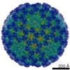 6969MC 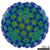 6968C  5zvsC M: map data used to model this data C: citing same article ( |
|---|---|
| Similar structure data |

-Links
 PDBj
PDBj


- Assembly
| Deposited unit | 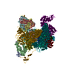
|
|---|---|
| 1 | x 60
|
| 2 |
|
| 3 | x 5
|
| 4 | x 6
|
| 5 | 
|
| Symmetry | Point symmetry: (Schoenflies symbol: I (icosahedral)) |
-Components
-Protein , 5 types, 25 molecules lbfdhjnprtBDFHJLNPRTUVWXY
| #1: Protein | Mass: 29844.648 Da / Num. of mol.: 10 Source method: isolated from a genetically manipulated source Source: (gene. exp.) Grass carp reovirus / Production host:  Ctenopharyngodon idella (grass carp) / References: UniProt: Q8JU63 Ctenopharyngodon idella (grass carp) / References: UniProt: Q8JU63#3: Protein | Mass: 64362.086 Da / Num. of mol.: 10 Source method: isolated from a genetically manipulated source Source: (gene. exp.) Grass carp reovirus / Production host: Ctenopharyngodon idella (grass carp) / References: UniProt: Q8JU67#4: Protein | Mass: 44606.535 Da / Num. of mol.: 2 Source method: isolated from a genetically manipulated source Source: (gene. exp.) Grass carp reovirus / Production host: Ctenopharyngodon idella (grass carp) / References: UniProt: Q8JU64#5: Protein | | Mass: 141512.156 Da / Num. of mol.: 1 Source method: isolated from a genetically manipulated source Source: (gene. exp.) Grass carp reovirus / Production host: Ctenopharyngodon idella (grass carp) / References: UniProt: Q9E3W0#6: Protein | Mass: 132203.312 Da / Num. of mol.: 2 Source method: isolated from a genetically manipulated source Source: (gene. exp.) Grass carp reovirus / Production host: Ctenopharyngodon idella (grass carp) / References: UniProt: Q9E3V8 |
|---|
-Protein/peptide / Non-polymers , 2 types, 20 molecules ACEGIKMOQS
| #2: Protein/peptide | Mass: 4302.686 Da / Num. of mol.: 10 Source method: isolated from a genetically manipulated source Source: (gene. exp.) Grass carp reovirus / Production host: Ctenopharyngodon idella (grass carp) / References: UniProt: Q8JU67#7: Chemical | ChemComp-MYR /  Mass: 228.371 Da / Num. of mol.: 10 / Source method: obtained synthetically / Formula: C14H28O2 Mass: 228.371 Da / Num. of mol.: 10 / Source method: obtained synthetically / Formula: C14H28O2 |
|---|
-Details
| Has protein modification | Y |
|---|
-Experimental details
-Experiment
| Experiment | Method: ELECTRON MICROSCOPY |
|---|---|
| EM experiment | Aggregation state: PARTICLE / 3D reconstruction method: single particle reconstruction |
- Sample preparation
| Component | Name: Grass carp reovirus / Type: VIRUS / Entity ID: #1-#6 / Source: RECOMBINANT |
|---|---|
| Source (natural) | Organism: Grass carp reovirus |
| Source (recombinant) | Organism: Ctenopharyngodon idella (grass carp) |
| Details of virus | Empty: NO / Enveloped: NO / Isolate: STRAIN / Type: VIRION |
| Buffer solution | pH: 7.5 |
| Specimen | Embedding applied: NO / Shadowing applied: NO / Staining applied: NO / Vitrification applied: YES |
| Vitrification | Cryogen name: ETHANE |
- Electron microscopy imaging
| Experimental equipment |  Model: Talos Arctica / Image courtesy: FEI Company |
|---|---|
| Microscopy | Model: FEI TECNAI ARCTICA |
| Electron gun | Electron source:  FIELD EMISSION GUN / Accelerating voltage: 200 kV / Illumination mode: OTHER FIELD EMISSION GUN / Accelerating voltage: 200 kV / Illumination mode: OTHER |
| Electron lens | Mode: BRIGHT FIELD |
| Image recording | Electron dose: 25 e/Å2 / Film or detector model: FEI FALCON II (4k x 4k) |
- Processing
| Software | Name: PHENIX / Version: 1.13_2998: / Classification: refinement |
|---|---|
| EM software | Name: PHENIX / Category: model refinement |
| CTF correction | Type: PHASE FLIPPING AND AMPLITUDE CORRECTION |
| 3D reconstruction | Resolution: 3.3 Å / Resolution method: FSC 0.143 CUT-OFF / Num. of particles: 41000 / Symmetry type: POINT |
