Major capsid protein, N-terminal / Major capsid protein N-terminus / Major capsid protein, C-terminal / Major capsid protein, C-terminal domain superfamily / Large eukaryotic DNA virus major capsid protein / Group II dsDNA virus coat/capsid protein Similarity search - Domain/homology
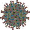
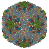
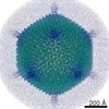
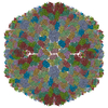
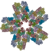
Journal: J Virol / Year: 2010 Title: Structural studies of the Sputnik virophage. Authors: Siyang Sun / Bernard La Scola / Valorie D Bowman / Christopher M Ryan / Julian P Whitelegge / Didier Raoult / Michael G Rossmann / Abstract: The virophage Sputnik is a satellite virus of the giant mimivirus and is the only satellite virus reported to date whose propagation adversely affects its host virus' production. Genome sequence ...The virophage Sputnik is a satellite virus of the giant mimivirus and is the only satellite virus reported to date whose propagation adversely affects its host virus' production. Genome sequence analysis showed that Sputnik has genes related to viruses infecting all three domains of life. Here, we report structural studies of Sputnik, which show that it is about 740 A in diameter, has a T=27 icosahedral capsid, and has a lipid membrane inside the protein shell. Structural analyses suggest that the major capsid protein of Sputnik is likely to have a double jelly-roll fold, although sequence alignments do not show any detectable similarity with other viral double jelly-roll capsid proteins. Hence, the origin of Sputnik's capsid might have been derived from other viruses prior to its association with mimivirus.
History
Deposition
Nov 4, 2009
Deposition site: RCSB / Processing site: RCSB
Revision 1.0
Nov 17, 2009
Provider: repository / Type: Initial release
Revision 1.1
Jul 13, 2011
Group: Version format compliance
Revision 1.2
Jan 24, 2018
Group: Data collection / Data processing / Structure summary Category: audit_author / em_image_scans / em_software / Item: _audit_author.name / _em_software.name
Revision 1.3
Jul 18, 2018
Group: Data collection / Category: em_software / Item: _em_software.image_processing_id
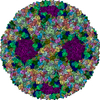
A: Major capsid protein B: Major capsid protein C: Major capsid protein D: Major capsid protein E: Major capsid protein F: Major capsid protein G: Major capsid protein H: Major capsid protein I: Major capsid protein J: Major capsid protein K: Major capsid protein L: Major capsid protein M: Major capsid protein
A: Major capsid protein B: Major capsid protein C: Major capsid protein D: Major capsid protein E: Major capsid protein F: Major capsid protein G: Major capsid protein H: Major capsid protein I: Major capsid protein J: Major capsid protein K: Major capsid protein L: Major capsid protein M: Major capsid protein
A: Major capsid protein B: Major capsid protein C: Major capsid protein D: Major capsid protein E: Major capsid protein F: Major capsid protein G: Major capsid protein H: Major capsid protein I: Major capsid protein J: Major capsid protein K: Major capsid protein L: Major capsid protein M: Major capsid protein
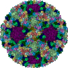
x 5
icosahedral pentamer
3.13 MDa, 65 polymers
Theoretical mass
Number of molelcules
Total (without water)
3,132,976
65
Polymers
3,132,976
65
Non-polymers
0
0
Water
0
Type
Name
Symmetry operation
Number
identity operation
1_555
x,y,z
1
point symmetry operation
4
4
A: Major capsid protein B: Major capsid protein C: Major capsid protein D: Major capsid protein E: Major capsid protein F: Major capsid protein G: Major capsid protein H: Major capsid protein I: Major capsid protein J: Major capsid protein K: Major capsid protein L: Major capsid protein M: Major capsid protein
x 6
icosahedral 23 hexamer
3.76 MDa, 78 polymers
Theoretical mass
Number of molelcules
Total (without water)
3,759,571
78
Polymers
3,759,571
78
Non-polymers
0
0
Water
0
Type
Name
Symmetry operation
Number
identity operation
1_555
x,y,z
1
point symmetry operation
5
5
Idetical with deposited unit in distinct coordinate
icosahedral asymmetric unit, std point frame
Type
Name
Symmetry operation
Number
transform to point frame
1
Symmetry
Point symmetry: (Schoenflies symbol: I (icosahedral))
Mass: 48199.625 Da / Num. of mol.: 13 / Source method: isolated from a natural source / Source: (natural) Acanthamoeba polyphaga (eukaryote) / References: UniProt: P30328
-
Experimental details
-
Experiment
Experiment
Method: ELECTRON MICROSCOPY
EM experiment
Aggregation state: PARTICLE / 3D reconstruction method: single particle reconstruction
-
Sample preparation
Component
Name: major capsid protein of sputnik
Details of virus
Host category: PROTOZOA / Isolate: SPECIES / Type: SATELLITE
Natural host
Organism: Acanthamoeba polyphaga
Buffer solution
Name: PBS / pH: 7.4 / Details: PBS
Specimen
Conc.: 1 mg/ml / Embedding applied: NO / Shadowing applied: NO / Staining applied: NO / Vitrification applied: YES
Vitrification
Instrument: HOMEMADE PLUNGER / Cryogen name: ETHANE / Details: flash-frozen on holey grids in liquid ethane
-
Electron microscopy imaging
Microscopy
Model: FEI/PHILIPS CM200FEG
Electron gun
Electron source: FIELD EMISSION GUN / Accelerating voltage: 200 kV / Illumination mode: FLOOD BEAM
Electron lens
Mode: BRIGHT FIELD / Nominal magnification: 38000 X / Calibrated magnification: 39190 X / Nominal defocus max: 3582 nm / Nominal defocus min: 767 nm / Cs: 2 mm
Specimen holder
Temperature: 70 K
Image recording
Electron dose: 20 e/Å2 / Film or detector model: GENERIC FILM
Radiation
Protocol: SINGLE WAVELENGTH / Monochromatic (M) / Laue (L): M / Scattering type: x-ray
Radiation wavelength
Relative weight: 1
-
Processing
EM software
ID
Name
Category
1
EMfit
modelfitting
2
EMAN
3Dreconstruction
CTF correction
Details: CTF parameters were calculated for particles in each micrograph.
Symmetry
Point symmetry: I (icosahedral)
3D reconstruction
Method: projection matching / Resolution: 10.6 Å / Num. of particles: 6780 / Nominal pixel size: 1.62 Å / Actual pixel size: 1.62 Å / Details: EMAN / Symmetry type: POINT
Atomic model building
Protocol: RIGID BODY FIT / Space: REAL / Target criteria: Sumf / Details: REFINEMENT PROTOCOL--rigid body
In the structure databanks used in Yorodumi, some data are registered as the other names, "COVID-19 virus" and "2019-nCoV". Here are the details of the virus and the list of structure data.
Jan 31, 2019. EMDB accession codes are about to change! (news from PDBe EMDB page)
EMDB accession codes are about to change! (news from PDBe EMDB page)
The allocation of 4 digits for EMDB accession codes will soon come to an end. Whilst these codes will remain in use, new EMDB accession codes will include an additional digit and will expand incrementally as the available range of codes is exhausted. The current 4-digit format prefixed with “EMD-” (i.e. EMD-XXXX) will advance to a 5-digit format (i.e. EMD-XXXXX), and so on. It is currently estimated that the 4-digit codes will be depleted around Spring 2019, at which point the 5-digit format will come into force.
The EM Navigator/Yorodumi systems omit the EMD- prefix.
Related info.:Q: What is EMD? / ID/Accession-code notation in Yorodumi/EM Navigator
Yorodumi is a browser for structure data from EMDB, PDB, SASBDB, etc.
This page is also the successor to EM Navigator detail page, and also detail information page/front-end page for Omokage search.
The word "yorodu" (or yorozu) is an old Japanese word meaning "ten thousand". "mi" (miru) is to see.
Related info.:EMDB / PDB / SASBDB / Comparison of 3 databanks / Yorodumi Search / Aug 31, 2016. New EM Navigator & Yorodumi / Yorodumi Papers / Jmol/JSmol / Function and homology information / Changes in new EM Navigator and Yorodumi
 Movie
Movie Controller

 Yorodumi
 Open data
 Basic information
 Components
 Keywords
 Function and homology information
 Acanthamoeba polyphaga (eukaryote)
 Authors
 Citation
 Structure visualization
 Downloads & links
 Other downloads
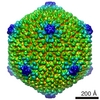


 PDBj
PDBj

 Assembly


 Sample preparation
 Electron microscopy imaging
 FIELD EMISSION GUN / Accelerating voltage: 200 kV / Illumination mode: FLOOD BEAM
 Processing
