 Movie
Movie Controller


+ Open data

- Basic information
| Entry | Database: PDB / ID: 5wda | ||||||||||||
|---|---|---|---|---|---|---|---|---|---|---|---|---|---|
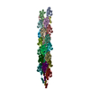
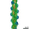
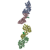
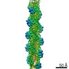
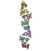
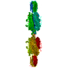
| Title | Structure of the PulG pseudopilus | ||||||||||||
 Components Components | General secretion pathway protein G | ||||||||||||
 Keywords Keywords | PROTEIN TRANSPORT / helical polymer / bacterial secretion / cryo-EM | ||||||||||||
| Function / homology |  Function and homology information Function and homology informationprotein secretion by the type II secretion system / type II protein secretion system complex / membrane => GO:0016020 / plasma membrane Similarity search - Function | ||||||||||||
| Biological species |  Klebsiella oxytoca (bacteria) Klebsiella oxytoca (bacteria) | ||||||||||||
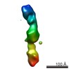
| Method | ELECTRON MICROSCOPY / helical reconstruction / cryo EM / Resolution: 5 Å | ||||||||||||
 Authors Authors | Lopez-Castilla, A. / Thomassin, J.L. / Bardiaux, B. / Zheng, W. / Nivaskumar, M. / Yu, X. / Nilges, M. / Egelman, E.H. / Izadi-Pruneyre, N. / Francetic, O. | ||||||||||||
| Funding support |  United States, United States,  France, European Union, 3items France, European Union, 3items
| ||||||||||||
 Citation Citation | Journal: Nat Microbiol / Year: 2017 Title: Structure of the calcium-dependent type 2 secretion pseudopilus. Authors: Aracelys López-Castilla / Jenny-Lee Thomassin / Benjamin Bardiaux / Weili Zheng / Mangayarkarasi Nivaskumar / Xiong Yu / Michael Nilges / Edward H Egelman / Nadia Izadi-Pruneyre / Olivera Francetic / Abstract: Many Gram-negative bacteria use type 2 secretion systems (T2SSs) to secrete proteins involved in virulence and adaptation. Transport of folded proteins via T2SS nanomachines requires the assembly of ...Many Gram-negative bacteria use type 2 secretion systems (T2SSs) to secrete proteins involved in virulence and adaptation. Transport of folded proteins via T2SS nanomachines requires the assembly of inner membrane-anchored fibres called pseudopili. Although efficient pseudopilus assembly is essential for protein secretion, structure-based functional analyses are required to unravel the mechanistic link between these processes. Here, we report an atomic model for a T2SS pseudopilus from Klebsiella oxytoca, obtained by fitting the NMR structure of its calcium-bound subunit PulG into the ~5-Å-resolution cryo-electron microscopy reconstruction of assembled fibres. This structure reveals the comprehensive network of inter-subunit contacts and unexpected features, including a disordered central region of the PulG helical stem, and highly flexible C-terminal residues on the fibre surface. NMR, mutagenesis and functional analyses highlight the key role of calcium in PulG folding and stability. Fibre disassembly in the absence of calcium provides a basis for pseudopilus length control, essential for protein secretion, and supports the Archimedes screw model for the type 2 secretion mechanism. | ||||||||||||
| History |
|
- Structure visualization
| Movie |
Movie viewer |
|---|---|
| Structure viewer | Molecule: MolmilJmol/JSmol |
- Downloads & links
-Download
| PDBx/mmCIF format | 5wda.cif.gz | 999.8 KB | Display | PDBx/mmCIF format |
|---|---|---|---|---|
| PDB format | pdb5wda.ent.gz | 873.9 KB | Display | PDB format |
| PDBx/mmJSON format | 5wda.json.gz | Tree view | PDBx/mmJSON format | |
| Others |  Other downloads Other downloads |
-Validation report
| Summary document | 5wda_validation.pdf.gz | 862.5 KB | Display | wwPDB validaton report |
|---|---|---|---|---|
| Full document | 5wda_full_validation.pdf.gz | 897.1 KB | Display | |
| Data in XML | 5wda_validation.xml.gz | 60.9 KB | Display | |
| Data in CIF | 5wda_validation.cif.gz | 103 KB | Display | |
| Arichive directory | https://data.pdbj.org/pub/pdb/validation_reports/wd/5wdaftp://data.pdbj.org/pub/pdb/validation_reports/wd/5wda | HTTPS FTP |
-Related structure data
| Related structure data |  8812MC 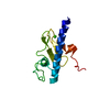 5o2yC C: citing same article ( M: map data used to model this data |
|---|---|
| Similar structure data |

-Links
 PDBj
PDBj
- Assembly
| Deposited unit | 
|
|---|---|
| 1 |
|
-Components
| #1: Protein | Mass: 14525.482 Da / Num. of mol.: 25 / Fragment: UNP residues 7-140 Source method: isolated from a genetically manipulated source Source: (gene. exp.) Klebsiella oxytoca (bacteria) / Gene: pulG, AB185_31145, SAMEA2273639_02747 / Production host: #2: Chemical | ChemComp-CA /   Mass: 40.078 Da / Num. of mol.: 25 / Source method: obtained synthetically / Formula: Ca Mass: 40.078 Da / Num. of mol.: 25 / Source method: obtained synthetically / Formula: Ca |
|---|
-Experimental details
-Experiment
| Experiment | Method: ELECTRON MICROSCOPY |
|---|---|
| EM experiment | Aggregation state: FILAMENT / 3D reconstruction method: helical reconstruction |
- Sample preparation
| Component | Name: PulG pseudopilus / Type: ORGANELLE OR CELLULAR COMPONENT / Entity ID: all / Source: RECOMBINANT |
|---|---|
| Molecular weight | Experimental value: NO |
| Source (natural) | Organism: Klebsiella oxytoca (bacteria) |
| Source (recombinant) | Organism: |
| Buffer solution | pH: 7.5 |
| Specimen | Embedding applied: NO / Shadowing applied: NO / Staining applied: NO / Vitrification applied: YES |
| Vitrification | Instrument: FEI VITROBOT MARK IV / Cryogen name: ETHANE |
- Electron microscopy imaging
| Experimental equipment |  Model: Titan Krios / Image courtesy: FEI Company |
|---|---|
| Microscopy | Model: FEI TITAN KRIOS |
| Electron gun | Electron source:  FIELD EMISSION GUN / Accelerating voltage: 300 kV / Illumination mode: FLOOD BEAM FIELD EMISSION GUN / Accelerating voltage: 300 kV / Illumination mode: FLOOD BEAM |
| Electron lens | Mode: BRIGHT FIELD |
| Image recording | Electron dose: 20 e/Å2 / Detector mode: INTEGRATING / Film or detector model: FEI FALCON II (4k x 4k) / Num. of grids imaged: 1 / Num. of real images: 1819 |
- Processing
| Software | Name: PHENIX / Version: 1.11.1_2575: / Classification: refinement | ||||||||||||||||||||||||
|---|---|---|---|---|---|---|---|---|---|---|---|---|---|---|---|---|---|---|---|---|---|---|---|---|---|
| EM software |
| ||||||||||||||||||||||||
| CTF correction | Type: PHASE FLIPPING AND AMPLITUDE CORRECTION | ||||||||||||||||||||||||
| Helical symmerty | Angular rotation/subunit: 83.2 ° / Axial rise/subunit: 10.2 Å / Axial symmetry: C1 | ||||||||||||||||||||||||
| 3D reconstruction | Resolution: 5 Å / Resolution method: FSC 0.143 CUT-OFF / Num. of particles: 85619 / Algorithm: BACK PROJECTION / Symmetry type: HELICAL | ||||||||||||||||||||||||
| Refine LS restraints |
|
