 Movie
Movie Controller

[English] 日本語
 Yorodumi
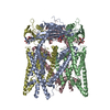
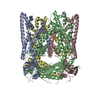
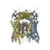
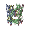
- PDB-5v4s: CryoEM Structure of a Prokaryotic Cyclic Nucleotide-Gated Ion Channel -
+ Open data

- Basic information
| Entry | Database: PDB / ID: 5v4s | |||||||||||||||
|---|---|---|---|---|---|---|---|---|---|---|---|---|---|---|---|---|
| Title | CryoEM Structure of a Prokaryotic Cyclic Nucleotide-Gated Ion Channel | |||||||||||||||
 Components Components | Transporter, cation channel family / cyclic nucleotide-binding domain multi-domain protein | |||||||||||||||
 Keywords Keywords | TRANSPORT PROTEIN / Ion channel / cyclic nucleotide / allostery / vision / olfaction | |||||||||||||||
| Function / homology |  Function and homology information Function and homology informationHCN channel complex / regulation of membrane depolarization / voltage-gated potassium channel activity / sodium ion transmembrane transport Similarity search - Function | |||||||||||||||
| Biological species |  Leptospira licerasiae serovar Varillal str. VAR 010 (bacteria) Leptospira licerasiae serovar Varillal str. VAR 010 (bacteria) | |||||||||||||||
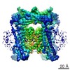
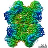
| Method | ELECTRON MICROSCOPY / single particle reconstruction / cryo EM / Resolution: 4.2 Å | |||||||||||||||
 Authors Authors | James, Z.M. / Borst, A.J. / Haitin, Y. / Frenz, B. / DiMaio, F. / Zagotta, W.N. / Veesler, D. | |||||||||||||||
| Funding support |  United States, 4items United States, 4items
| |||||||||||||||
 Citation Citation | Journal: Proc Natl Acad Sci U S A / Year: 2017 Title: CryoEM structure of a prokaryotic cyclic nucleotide-gated ion channel. Authors: Zachary M James / Andrew J Borst / Yoni Haitin / Brandon Frenz / Frank DiMaio / William N Zagotta / David Veesler /  Abstract: Cyclic nucleotide-gated (CNG) and hyperpolarization-activated cyclic nucleotide-regulated (HCN) ion channels play crucial physiological roles in phototransduction, olfaction, and cardiac pace making. ...Cyclic nucleotide-gated (CNG) and hyperpolarization-activated cyclic nucleotide-regulated (HCN) ion channels play crucial physiological roles in phototransduction, olfaction, and cardiac pace making. These channels are characterized by the presence of a carboxyl-terminal cyclic nucleotide-binding domain (CNBD) that connects to the channel pore via a C-linker domain. Although cyclic nucleotide binding has been shown to promote CNG and HCN channel opening, the precise mechanism underlying gating remains poorly understood. Here we used cryoEM to determine the structure of the intact LliK CNG channel isolated from -which shares sequence similarity to eukaryotic CNG and HCN channels-in the presence of a saturating concentration of cAMP. A short S4-S5 linker connects nearby voltage-sensing and pore domains to produce a non-domain-swapped transmembrane architecture, which appears to be a hallmark of this channel family. We also observe major conformational changes of the LliK C-linkers and CNBDs relative to the crystal structures of isolated C-linker/CNBD fragments and the cryoEM structures of related CNG, HCN, and KCNH channels. The conformation of our LliK structure may represent a functional state of this channel family not captured in previous studies. | |||||||||||||||
| History |
|
- Structure visualization
| Movie |
Movie viewer |
|---|---|
| Structure viewer | Molecule: MolmilJmol/JSmol |
- Downloads & links
-Download
| PDBx/mmCIF format | 5v4s.cif.gz | 275.8 KB | Display | PDBx/mmCIF format |
|---|---|---|---|---|
| PDB format | pdb5v4s.ent.gz | 212 KB | Display | PDB format |
| PDBx/mmJSON format | 5v4s.json.gz | Tree view | PDBx/mmJSON format | |
| Others |  Other downloads Other downloads |
-Validation report
| Arichive directory | https://data.pdbj.org/pub/pdb/validation_reports/v4/5v4sftp://data.pdbj.org/pub/pdb/validation_reports/v4/5v4s | HTTPS FTP |
|---|
-Related structure data
| Related structure data |  8632MC  8633MC M: map data used to model this data C: citing same article ( |
|---|---|
| Similar structure data |


-Links
 PDBj
PDBj


- Assembly
| Deposited unit | 
|
|---|---|
| 1 |
|
-Components
| #1: Protein | Mass: 53464.785 Da / Num. of mol.: 4 Source method: isolated from a genetically manipulated source Source: (gene. exp.) Leptospira licerasiae serovar Varillal str. VAR 010 (bacteria)Gene: LEP1GSC185_1946 / Production host: |
|---|
-Experimental details
-Experiment
| Experiment | Method: ELECTRON MICROSCOPY |
|---|---|
| EM experiment | Aggregation state: PARTICLE / 3D reconstruction method: single particle reconstruction |
- Sample preparation
| Component | Name: LliK / Type: COMPLEX / Entity ID: all / Source: RECOMBINANT | ||||||||||||||||||||||||||||||
|---|---|---|---|---|---|---|---|---|---|---|---|---|---|---|---|---|---|---|---|---|---|---|---|---|---|---|---|---|---|---|---|
| Molecular weight | Value: 0.2 MDa / Experimental value: NO | ||||||||||||||||||||||||||||||
| Source (natural) | Organism: Leptospira licerasiae serovar Varillal str. VAR 010 (bacteria) | ||||||||||||||||||||||||||||||
| Source (recombinant) | Organism: | ||||||||||||||||||||||||||||||
| Buffer solution | pH: 7.9 | ||||||||||||||||||||||||||||||
| Buffer component |
| ||||||||||||||||||||||||||||||
| Specimen | Conc.: 0.9 mg/ml / Embedding applied: NO / Shadowing applied: NO / Staining applied: NO / Vitrification applied: YES | ||||||||||||||||||||||||||||||
| Specimen support | Grid material: COPPER / Grid mesh size: 400 divisions/in. / Grid type: Quantifoil R1.2/1.3 | ||||||||||||||||||||||||||||||
| Vitrification | Cryogen name: ETHANE |
- Electron microscopy imaging
| Experimental equipment |  Model: Titan Krios / Image courtesy: FEI Company |
|---|---|
| Microscopy | Model: FEI TITAN KRIOS |
| Electron gun | Electron source:  FIELD EMISSION GUN / Accelerating voltage: 300 kV / Illumination mode: FLOOD BEAM FIELD EMISSION GUN / Accelerating voltage: 300 kV / Illumination mode: FLOOD BEAM |
| Electron lens | Mode: BRIGHT FIELD |
| Image recording | Electron dose: 1.4 e/Å2 / Detector mode: COUNTING / Film or detector model: GATAN K2 SUMMIT (4k x 4k) |
- Processing
| EM software |
| ||||||||||||||||||||||||||||||||||||||||
|---|---|---|---|---|---|---|---|---|---|---|---|---|---|---|---|---|---|---|---|---|---|---|---|---|---|---|---|---|---|---|---|---|---|---|---|---|---|---|---|---|---|
| CTF correction | Type: PHASE FLIPPING AND AMPLITUDE CORRECTION | ||||||||||||||||||||||||||||||||||||||||
| Particle selection | Num. of particles selected: 379000 | ||||||||||||||||||||||||||||||||||||||||
| Symmetry | Point symmetry: C4 (4 fold cyclic) | ||||||||||||||||||||||||||||||||||||||||
| 3D reconstruction | Resolution: 4.2 Å / Resolution method: FSC 0.143 CUT-OFF / Num. of particles: 18737 / Symmetry type: POINT | ||||||||||||||||||||||||||||||||||||||||
| Atomic model building | Protocol: OTHER / Space: REAL |
