 Movie
Movie Controller

[English] 日本語
 Yorodumi
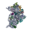
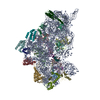
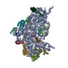
- PDB-5uz4: The cryo-EM structure of YjeQ bound to the 30S subunit suggests a... -
+ Open data

- Basic information
| Entry | Database: PDB / ID: 5uz4 | ||||||
|---|---|---|---|---|---|---|---|
| Title | The cryo-EM structure of YjeQ bound to the 30S subunit suggests a fidelity checkpoint function for this protein in ribosome assembly | ||||||
 Components Components |
| ||||||
 Keywords Keywords | RIBOSOME/Hydrolase / Ribosome assembly / 30S subunit / YjeQ protein / RsgA protein / RIBOSOME-Hydrolase complex | ||||||
| Function / homology |  Function and homology information Function and homology informationguanosine tetraphosphate binding / transcription antitermination factor activity, RNA binding / ornithine decarboxylase inhibitor activity / Hydrolases; Acting on acid anhydrides; In phosphorus-containing anhydrides / four-way junction DNA binding / negative regulation of translational initiation / mRNA regulatory element binding translation repressor activity / regulation of DNA-templated transcription elongation / transcription elongation factor complex / transcription antitermination ...guanosine tetraphosphate binding / transcription antitermination factor activity, RNA binding / ornithine decarboxylase inhibitor activity / Hydrolases; Acting on acid anhydrides; In phosphorus-containing anhydrides / four-way junction DNA binding / negative regulation of translational initiation / mRNA regulatory element binding translation repressor activity / regulation of DNA-templated transcription elongation / transcription elongation factor complex / transcription antitermination / DNA endonuclease activity / DNA-templated transcription termination / maintenance of translational fidelity / mRNA 5'-UTR binding / GDP binding / regulation of translation / ribosomal small subunit assembly / ribosome biogenesis / ribosomal small subunit biogenesis / small ribosomal subunit / small ribosomal subunit rRNA binding / cytosolic small ribosomal subunit / cytoplasmic translation / tRNA binding / negative regulation of translation / rRNA binding / structural constituent of ribosome / ribosome / translation / ribonucleoprotein complex / response to antibiotic / hydrolase activity / mRNA binding / GTPase activity / GTP binding / RNA binding / zinc ion binding / membrane / metal ion binding / cytoplasm / cytosol Similarity search - Function | ||||||
| Biological species |  | ||||||
| Method | ELECTRON MICROSCOPY / single particle reconstruction / cryo EM / Resolution: 5.8 Å | ||||||
 Authors Authors | Razi, A. / Guarne, A. / Ortega, J. | ||||||
| Funding support |  Canada, 1items Canada, 1items
| ||||||
 Citation Citation | Journal: Proc Natl Acad Sci U S A / Year: 2017 Title: The cryo-EM structure of YjeQ bound to the 30S subunit suggests a fidelity checkpoint function for this protein in ribosome assembly. Authors: Aida Razi / Alba Guarné / Joaquin Ortega / Abstract: Recent work suggests that bacterial YjeQ (RsgA) participates in the late stages of assembly of the 30S subunit and aids the assembly of the decoding center but also binds the mature 30S subunit with ...Recent work suggests that bacterial YjeQ (RsgA) participates in the late stages of assembly of the 30S subunit and aids the assembly of the decoding center but also binds the mature 30S subunit with high affinity. To determine the function and mechanisms of YjeQ in the context of the mature subunit, we determined the cryo-EM structure of the fully assembled 30S subunit in complex with YjeQ at 5.8-Å resolution. We found that binding of YjeQ stabilizes helix 44 into a conformation similar to that adopted by the subunit during proofreading. This finding indicates that, along with acting as an assembly factor, YjeQ has a role as a checkpoint protein, consisting of testing the proofreading ability of the 30S subunit. The structure also informs the mechanism by which YjeQ implements the release from the 30S subunit of a second assembly factor, called RbfA. Finally, it reveals how the 30S subunit stimulates YjeQ GTPase activity and leads to release of the protein. Checkpoint functions have been described for eukaryotic ribosome assembly factors; however, this work describes an example of a bacterial assembly factor that tests a specific translation mechanism of the 30S subunit. | ||||||
| History |
|
- Structure visualization
| Movie |
Movie viewer |
|---|---|
| Structure viewer | Molecule: MolmilJmol/JSmol |
- Downloads & links
-Download
| PDBx/mmCIF format | 5uz4.cif.gz | 1.2 MB | Display | PDBx/mmCIF format |
|---|---|---|---|---|
| PDB format | pdb5uz4.ent.gz | 923.8 KB | Display | PDB format |
| PDBx/mmJSON format | 5uz4.json.gz | Tree view | PDBx/mmJSON format | |
| Others |  Other downloads Other downloads |
-Validation report
| Arichive directory | https://data.pdbj.org/pub/pdb/validation_reports/uz/5uz4ftp://data.pdbj.org/pub/pdb/validation_reports/uz/5uz4 | HTTPS FTP |
|---|
-Related structure data
| Related structure data |  8621MC 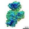 8626C 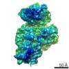 8627C 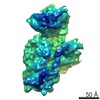 8628C C: citing same article ( M: map data used to model this data |
|---|---|
| Similar structure data |


-Links
 PDBj
PDBj


- Assembly
| Deposited unit | 
|
|---|---|
| 1 |
|
-Components
-30S ribosomal protein ... , 19 types, 19 molecules CDEFGHIJKLMNOPQRSTB
| #2: Protein | Mass: 26031.316 Da / Num. of mol.: 1 / Source method: isolated from a natural source / Source: (natural) |
|---|---|
| #3: Protein | Mass: 23514.199 Da / Num. of mol.: 1 / Source method: isolated from a natural source / Source: (natural) |
| #4: Protein | Mass: 17629.398 Da / Num. of mol.: 1 / Source method: isolated from a natural source / Source: (natural) |
| #5: Protein | Mass: 15211.058 Da / Num. of mol.: 1 / Source method: isolated from a natural source / Source: (natural) |
| #6: Protein | Mass: 20055.156 Da / Num. of mol.: 1 / Source method: isolated from a natural source / Source: (natural) |
| #7: Protein | Mass: 14146.557 Da / Num. of mol.: 1 / Source method: isolated from a natural source / Source: (natural) |
| #8: Protein | Mass: 14886.270 Da / Num. of mol.: 1 / Source method: isolated from a natural source / Source: (natural) |
| #9: Protein | Mass: 11755.597 Da / Num. of mol.: 1 / Source method: isolated from a natural source / Source: (natural) |
| #10: Protein | Mass: 13870.975 Da / Num. of mol.: 1 / Source method: isolated from a natural source / Source: (natural) |
| #11: Protein | Mass: 13768.157 Da / Num. of mol.: 1 / Source method: isolated from a natural source / Source: (natural) |
| #12: Protein | Mass: 13128.467 Da / Num. of mol.: 1 / Source method: isolated from a natural source / Source: (natural) |
| #13: Protein | Mass: 11606.560 Da / Num. of mol.: 1 / Source method: isolated from a natural source / Source: (natural) |
| #14: Protein | Mass: 10319.882 Da / Num. of mol.: 1 / Source method: isolated from a natural source / Source: (natural) |
| #15: Protein | Mass: 9207.572 Da / Num. of mol.: 1 / Source method: isolated from a natural source / Source: (natural) |
| #16: Protein | Mass: 9724.491 Da / Num. of mol.: 1 / Source method: isolated from a natural source / Source: (natural) |
| #17: Protein | Mass: 9005.472 Da / Num. of mol.: 1 / Source method: isolated from a natural source / Source: (natural) |
| #18: Protein | Mass: 10455.355 Da / Num. of mol.: 1 / Source method: isolated from a natural source / Source: (natural) |
| #19: Protein | Mass: 9708.464 Da / Num. of mol.: 1 / Source method: isolated from a natural source / Source: (natural) |
| #20: Protein | Mass: 26781.670 Da / Num. of mol.: 1 / Source method: isolated from a natural source / Source: (natural) |
-RNA chain / Protein , 2 types, 2 molecules AZ
| #1: RNA chain | Mass: 494971.188 Da / Num. of mol.: 1 / Source method: isolated from a natural source / Source: (natural) |
|---|---|
| #21: Protein | Mass: 37345.172 Da / Num. of mol.: 1 Source method: isolated from a genetically manipulated source Source: (gene. exp.) References: UniProt: P39286, Hydrolases; Acting on acid anhydrides; In phosphorus-containing anhydrides |
-Non-polymers , 2 types, 2 molecules 

| #22: Chemical | ChemComp-ZN /  Mass: 65.409 Da / Num. of mol.: 1 / Source method: obtained synthetically / Formula: Zn Mass: 65.409 Da / Num. of mol.: 1 / Source method: obtained synthetically / Formula: Zn |
|---|---|
| #23: Chemical | ChemComp-GGM /  Mass: 655.343 Da / Num. of mol.: 1 / Source method: obtained synthetically / Formula: C18H24N7O14P3 Mass: 655.343 Da / Num. of mol.: 1 / Source method: obtained synthetically / Formula: C18H24N7O14P3 |
-Details
| Has protein modification | Y |
|---|
-Experimental details
-Experiment
| Experiment | Method: ELECTRON MICROSCOPY |
|---|---|
| EM experiment | Aggregation state: PARTICLE / 3D reconstruction method: single particle reconstruction |
- Sample preparation
| Component | Name: Structure of the 30S subunit in complex with YjeQ GTPase Type: COMPLEX / Entity ID: #1-#21 / Source: MULTIPLE SOURCES |
|---|---|
| Source (natural) | Organism: |
| Buffer solution | pH: 7.5 |
| Specimen | Embedding applied: NO / Shadowing applied: NO / Staining applied: NO / Vitrification applied: YES |
| Specimen support | Grid material: COPPER / Grid mesh size: 200 divisions/in. / Grid type: C-flat CFT-222C |
| Vitrification | Instrument: FEI VITROBOT MARK III / Cryogen name: ETHANE / Humidity: 100 % / Chamber temperature: 298 K |
- Electron microscopy imaging
| Experimental equipment |  Model: Tecnai F20 / Image courtesy: FEI Company |
|---|---|
| Microscopy | Model: FEI TECNAI F20 |
| Electron gun | Electron source:  FIELD EMISSION GUN / Accelerating voltage: 200 kV / Illumination mode: FLOOD BEAM FIELD EMISSION GUN / Accelerating voltage: 200 kV / Illumination mode: FLOOD BEAM |
| Electron lens | Mode: BRIGHT FIELD / Nominal magnification: 25000 X / Calibrated magnification: 34482 X / Nominal defocus max: 3000 nm / Nominal defocus min: 500 nm / Cs: 2 mm / C2 aperture diameter: 50 µm / Alignment procedure: COMA FREE |
| Specimen holder | Cryogen: NITROGEN Specimen holder model: GATAN 626 SINGLE TILT LIQUID NITROGEN CRYO TRANSFER HOLDER |
| Image recording | Average exposure time: 0.5 sec. / Electron dose: 1 e/Å2 / Detector mode: COUNTING / Film or detector model: GATAN K2 SUMMIT (4k x 4k) |
| Image scans | Movie frames/image: 30 / Used frames/image: 1-20 |
- Processing
| EM software |
| ||||||||||||||||||||
|---|---|---|---|---|---|---|---|---|---|---|---|---|---|---|---|---|---|---|---|---|---|
| CTF correction | Type: PHASE FLIPPING AND AMPLITUDE CORRECTION | ||||||||||||||||||||
| Particle selection | Num. of particles selected: 487018 | ||||||||||||||||||||
| 3D reconstruction | Resolution: 5.8 Å / Resolution method: FSC 0.143 CUT-OFF / Num. of particles: 130462 / Symmetry type: POINT | ||||||||||||||||||||
| Atomic model building | Protocol: OTHER |
