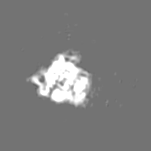
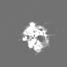
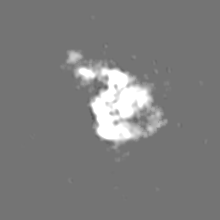
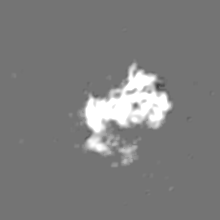
- EMDB-8628: Structure of the 30S subunit, subclass II -
+
Open data
ID or keywords:
Loading...
-
Basic information
Entry
Database: EMDB / ID: EMD-8628
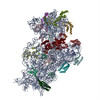
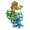
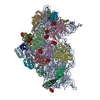
Title
Structure of the 30S subunit, subclass II
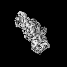
Map data
Sample
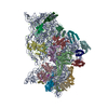
Complex: 30S Consensus subunit, subclass II
Function / homology
Function and homology information
guanosine tetraphosphate binding / transcription antitermination factor activity, RNA binding / ornithine decarboxylase inhibitor activity / Hydrolases; Acting on acid anhydrides; In phosphorus-containing anhydrides / four-way junction DNA binding / negative regulation of translational initiation / mRNA regulatory element binding translation repressor activity / regulation of DNA-templated transcription elongation / transcription elongation factor complex / transcription antitermination ...guanosine tetraphosphate binding / transcription antitermination factor activity, RNA binding / ornithine decarboxylase inhibitor activity / Hydrolases; Acting on acid anhydrides; In phosphorus-containing anhydrides / four-way junction DNA binding / negative regulation of translational initiation / mRNA regulatory element binding translation repressor activity / regulation of DNA-templated transcription elongation / transcription elongation factor complex / transcription antitermination / DNA endonuclease activity / DNA-templated transcription termination / maintenance of translational fidelity / mRNA 5'-UTR binding / GDP binding / regulation of translation / ribosomal small subunit assembly / ribosome biogenesis / ribosomal small subunit biogenesis / small ribosomal subunit / small ribosomal subunit rRNA binding / cytosolic small ribosomal subunit / cytoplasmic translation / tRNA binding / negative regulation of translation / rRNA binding / structural constituent of ribosome / ribosome / translation / ribonucleoprotein complex / response to antibiotic / hydrolase activity / mRNA binding / GTPase activity / GTP binding / RNA binding / zinc ion binding / membrane / metal ion binding / cytoplasm / cytosol Similarity search - Function
Ribosome biogenesis GTPase RsgA / RsgA GTPase domain / RsgA GTPase / EngC GTPase domain profile. / Circularly permuted (CP)-type guanine nucleotide-binding (G) domain / Circularly permuted (CP)-type guanine nucleotide-binding (G) domain profile. / Ribosomal protein S14, bacterial/plastid / Ribosomal protein S16, conserved site / Ribosomal protein S16 signature. / Ribosomal protein S6, conserved site ...Ribosome biogenesis GTPase RsgA / RsgA GTPase domain / RsgA GTPase / EngC GTPase domain profile. / Circularly permuted (CP)-type guanine nucleotide-binding (G) domain / Circularly permuted (CP)-type guanine nucleotide-binding (G) domain profile. / Ribosomal protein S14, bacterial/plastid / Ribosomal protein S16, conserved site / Ribosomal protein S16 signature. / Ribosomal protein S6, conserved site / Ribosomal protein S6 signature. / Ribosomal protein S3, bacterial-type / Ribosomal protein S13, bacterial-type / Ribosomal protein S19, bacterial-type / Ribosomal protein S7, bacterial/organellar-type / Ribosomal protein S11, bacterial-type / Ribosomal protein S20 / Ribosomal protein S20 superfamily / Ribosomal protein S20 / Ribosomal protein S4, bacterial-type / Ribosomal protein S5, bacterial-type / 30S ribosomal protein S17 / Ribosomal protein S6, plastid/chloroplast / Ribosomal protein S2, bacteria/mitochondria/plastid / Ribosomal protein S18, conserved site / Ribosomal protein S18 signature. / Ribosomal protein S9, bacterial/plastid / Ribosomal protein S16 / Ribosomal protein S16 domain superfamily / Ribosomal protein S16 / Ribosomal protein S15, bacterial-type / Ribosomal protein S6 / Ribosomal protein S6 / Ribosomal protein S6 superfamily / Ribosomal protein S12, bacterial-type / Translation elongation factor EF1B/ribosomal protein S6 / Ribosomal protein S18 / Ribosomal protein S18 / Ribosomal protein S18 superfamily / K Homology domain / K homology RNA-binding domain / Ribosomal protein S2 signature 2. / Ribosomal protein S3, conserved site / Ribosomal protein S3 signature. / Ribosomal protein S10, conserved site / Ribosomal protein S10 signature. / : / Ribosomal protein S14, conserved site / Ribosomal protein S14 signature. / Ribosomal protein S2 signature 1. / KH domain / Type-2 KH domain profile. / K Homology domain, type 2 / Ribosomal protein S3, C-terminal / Ribosomal protein S3, C-terminal domain / Ribosomal protein S3, C-terminal domain superfamily / Ribosomal protein S10 / Ribosomal protein S15/S19, conserved site / Ribosomal protein S19 signature. / Ribosomal protein S19/S15 / Ribosomal protein S19/S15, superfamily / Ribosomal protein S19 / Ribosomal protein S5, N-terminal, conserved site / Ribosomal protein S5 signature. / Ribosomal protein S7, conserved site / Ribosomal protein S7 signature. / K homology domain superfamily, prokaryotic type / Ribosomal protein S2, conserved site / : / Ribosomal protein S2 / Ribosomal protein S2, flavodoxin-like domain superfamily / Ribosomal protein S2 / Ribosomal protein S17, conserved site / Ribosomal protein S17 signature. / Ribosomal protein S5 / S5 double stranded RNA-binding domain profile. / Ribosomal protein S5, N-terminal / Ribosomal protein S5, C-terminal / Ribosomal protein S5, N-terminal domain / Ribosomal protein S13, conserved site / Ribosomal protein S13 signature. / Ribosomal protein S5, C-terminal domain / Ribosomal protein S13 / 30s ribosomal protein S13, C-terminal / Ribosomal protein S13/S18 / Ribosomal protein S13 family profile. / K homology domain-like, alpha/beta / Ribosomal protein S8 signature. / Ribosomal protein S4/S9 N-terminal domain / Ribosomal protein S14 / Ribosomal protein S14p/S29e / Ribosomal protein S4, conserved site / Ribosomal protein S4 signature. / Ribosomal protein S4/S9 N-terminal domain / Ribosomal protein S4/S9, N-terminal / Ribosomal protein S15 signature. / Ribosomal protein S4/S9 / Ribosomal protein S8 / Ribosomal protein S8 superfamily / Ribosomal protein S8 Similarity search - Domain/homology
Small ribosomal subunit protein bS20 / Small ribosomal subunit protein uS2 / Small ribosomal subunit protein uS9 / Small ribosomal subunit protein uS4 / Small ribosomal subunit protein uS11 / Small ribosomal subunit protein uS8 / Small ribosomal subunit protein uS14 / Small ribosomal subunit protein uS17 / Small ribosomal subunit protein uS3 / Small ribosomal subunit protein uS19 ...Small ribosomal subunit protein bS20 / Small ribosomal subunit protein uS2 / Small ribosomal subunit protein uS9 / Small ribosomal subunit protein uS4 / Small ribosomal subunit protein uS11 / Small ribosomal subunit protein uS8 / Small ribosomal subunit protein uS14 / Small ribosomal subunit protein uS17 / Small ribosomal subunit protein uS3 / Small ribosomal subunit protein uS19 / Small ribosomal subunit protein uS10 / Small ribosomal subunit protein uS12 / Small ribosomal subunit protein bS16 / Small ribosomal subunit protein bS18 / Small ribosomal subunit protein bS6 / Small ribosomal subunit protein uS7 / Small ribosomal subunit protein uS10 / Small ribosomal subunit protein uS13 / Small ribosomal subunit protein uS13 / Small ribosomal subunit protein bS16 / Small ribosomal subunit protein uS19 / Small ribosomal subunit protein bS20 / Small ribosomal subunit protein uS2 / Small ribosomal subunit protein uS3 / Small ribosomal subunit protein uS4 / Small ribosomal subunit protein uS5 / Small ribosomal subunit protein uS5 / Small ribosomal subunit protein uS15 / Small ribosomal subunit protein uS14 / Small ribosomal subunit biogenesis GTPase RsgA / Small ribosomal subunit protein uS15 Similarity search - Component
Biological species
Escherichia coli (E. coli)
Method
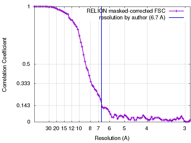
single particle reconstruction / cryo EM / Resolution: 6.7 Å
Journal: Proc Natl Acad Sci U S A / Year: 2017 Title: The cryo-EM structure of YjeQ bound to the 30S subunit suggests a fidelity checkpoint function for this protein in ribosome assembly. Authors: Aida Razi / Alba Guarné / Joaquin Ortega / Abstract: Recent work suggests that bacterial YjeQ (RsgA) participates in the late stages of assembly of the 30S subunit and aids the assembly of the decoding center but also binds the mature 30S subunit with ...Recent work suggests that bacterial YjeQ (RsgA) participates in the late stages of assembly of the 30S subunit and aids the assembly of the decoding center but also binds the mature 30S subunit with high affinity. To determine the function and mechanisms of YjeQ in the context of the mature subunit, we determined the cryo-EM structure of the fully assembled 30S subunit in complex with YjeQ at 5.8-Å resolution. We found that binding of YjeQ stabilizes helix 44 into a conformation similar to that adopted by the subunit during proofreading. This finding indicates that, along with acting as an assembly factor, YjeQ has a role as a checkpoint protein, consisting of testing the proofreading ability of the 30S subunit. The structure also informs the mechanism by which YjeQ implements the release from the 30S subunit of a second assembly factor, called RbfA. Finally, it reveals how the 30S subunit stimulates YjeQ GTPase activity and leads to release of the protein. Checkpoint functions have been described for eukaryotic ribosome assembly factors; however, this work describes an example of a bacterial assembly factor that tests a specific translation mechanism of the 30S subunit.
History
Deposition
Feb 27, 2017
-
Header (metadata) release
Mar 15, 2017
-
Map release
Apr 19, 2017
-
Update
Jul 18, 2018
-
Current status
Jul 18, 2018
Processing site: RCSB / Status: Released
-
Structure visualization
Movie
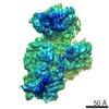
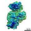
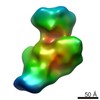
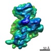
Surface view with section colored by density value
pH: 7.5 Details: 10 mM Tris-HCl at pH 7.5, 10 mM magnesium acetate, 150 mM NH4Cl, 3mM 2-mercaptoethanol and 2 mM GMP-PNP
Grid
Model: C-flat CFT-222C / Material: COPPER / Mesh: 200 / Support film - Material: CARBON / Support film - topology: HOLEY / Pretreatment - Type: GLOW DISCHARGE
Vitrification
Cryogen name: ETHANE / Chamber humidity: 100 % / Chamber temperature: 298 K / Instrument: FEI VITROBOT MARK III
-
Electron microscopy
Microscope
FEI TECNAI F20
Image recording
Film or detector model: GATAN K2 SUMMIT (4k x 4k) / Detector mode: COUNTING / Digitization - Frames/image: 1-20 / Average exposure time: 0.5 sec. / Average electron dose: 2.0 e/Å2
Electron beam
Acceleration voltage: 200 kV / Electron source: FIELD EMISSION GUN
In the structure databanks used in Yorodumi, some data are registered as the other names, "COVID-19 virus" and "2019-nCoV". Here are the details of the virus and the list of structure data.
Jan 31, 2019. EMDB accession codes are about to change! (news from PDBe EMDB page)
EMDB accession codes are about to change! (news from PDBe EMDB page)
The allocation of 4 digits for EMDB accession codes will soon come to an end. Whilst these codes will remain in use, new EMDB accession codes will include an additional digit and will expand incrementally as the available range of codes is exhausted. The current 4-digit format prefixed with “EMD-” (i.e. EMD-XXXX) will advance to a 5-digit format (i.e. EMD-XXXXX), and so on. It is currently estimated that the 4-digit codes will be depleted around Spring 2019, at which point the 5-digit format will come into force.
The EM Navigator/Yorodumi systems omit the EMD- prefix.
Related info.:Q: What is EMD? / ID/Accession-code notation in Yorodumi/EM Navigator
Yorodumi is a browser for structure data from EMDB, PDB, SASBDB, etc.
This page is also the successor to EM Navigator detail page, and also detail information page/front-end page for Omokage search.
The word "yorodu" (or yorozu) is an old Japanese word meaning "ten thousand". "mi" (miru) is to see.
Related info.:EMDB / PDB / SASBDB / Comparison of 3 databanks / Yorodumi Search / Aug 31, 2016. New EM Navigator & Yorodumi / Yorodumi Papers / Jmol/JSmol / Function and homology information / Changes in new EM Navigator and Yorodumi
 Movie
Movie Controller


 Open data
 Basic information
 Map data
 Sample
 Function and homology information
 Authors
 Citation
 Structure visualization
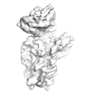
 Downloads & links
 emd_8628.png
 http://ftp.pdbj.org/pub/emdb/structures/EMD-8628


 Z (Sec.)
 Y (Row.)
 X (Col.)


 Sample components
 Processing
 Electron microscopy
 FIELD EMISSION GUN