+ Open data
- Basic information
| Entry | Database: PDB / ID: 5go9 | |||||||||
|---|---|---|---|---|---|---|---|---|---|---|
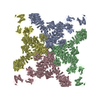
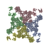
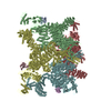
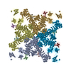
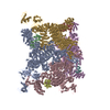
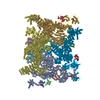
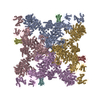
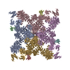
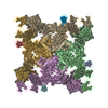
| Title | Cryo-EM structure of RyR2 in closed state | |||||||||
 Components Components | RyR2 | |||||||||
 Keywords Keywords | TRANSPORT PROTEIN / Membrane protein / Channel | |||||||||
| Biological species |  | |||||||||
| Method | ELECTRON MICROSCOPY / single particle reconstruction / cryo EM / Resolution: 4.4 Å | |||||||||
 Authors Authors | Peng, W. / Wu, J.P. / Yan, N. | |||||||||
| Funding support |  China, 2items China, 2items
| |||||||||
 Citation Citation |  Journal: Science / Year: 2016 Journal: Science / Year: 2016Title: Structural basis for the gating mechanism of the type 2 ryanodine receptor RyR2. Authors: Wei Peng / Huaizong Shen / Jianping Wu / Wenting Guo / Xiaojing Pan / Ruiwu Wang / S R Wayne Chen / Nieng Yan /   Abstract: RyR2 is a high-conductance intracellular calcium (Ca) channel that controls the release of Ca from the sarco(endo)plasmic reticulum of a variety of cells. Here, we report the structures of RyR2 from ...RyR2 is a high-conductance intracellular calcium (Ca) channel that controls the release of Ca from the sarco(endo)plasmic reticulum of a variety of cells. Here, we report the structures of RyR2 from porcine heart in both the open and closed states at near-atomic resolutions determined using single-particle electron cryomicroscopy. Structural comparison reveals a breathing motion of the overall cytoplasmic region resulted from the interdomain movements of amino-terminal domains (NTDs), Helical domains, and Handle domains, whereas almost no intradomain shifts are observed in these armadillo repeats-containing domains. Outward rotations of the Central domains, which integrate the conformational changes of the cytoplasmic region, lead to the dilation of the cytoplasmic gate through coupled motions. Our structural and mutational characterizations provide important insights into the gating and disease mechanism of RyRs. | |||||||||
| History |
|
- Structure visualization
| Movie |
 Movie viewer Movie viewer |
|---|---|
| Structure viewer | Molecule:  Molmil Molmil Jmol/JSmol Jmol/JSmol |
- Downloads & links
- Download
| PDBx/mmCIF format |  5go9.cif.gz 5go9.cif.gz | 2.6 MB | Display |  PDBx/mmCIF format PDBx/mmCIF format |
|---|---|---|---|---|
| PDB format |  pdb5go9.ent.gz pdb5go9.ent.gz | Display |  PDB format PDB format | |
| PDBx/mmJSON format |  5go9.json.gz 5go9.json.gz | Tree view |  PDBx/mmJSON format PDBx/mmJSON format | |
| Others |  Other downloads Other downloads |
-Validation report
| Arichive directory |  https://data.pdbj.org/pub/pdb/validation_reports/go/5go9 https://data.pdbj.org/pub/pdb/validation_reports/go/5go9 ftp://data.pdbj.org/pub/pdb/validation_reports/go/5go9 ftp://data.pdbj.org/pub/pdb/validation_reports/go/5go9 | HTTPS FTP |
|---|
-Related structure data
| Related structure data |  9528MC  9529C  5goaC M: map data used to model this data C: citing same article ( |
|---|---|
| Similar structure data |
- Links
Links
- Assembly
| Deposited unit | 
|
|---|---|
| 1 |
|
- Components
| #1: Protein | Mass: 564905.625 Da / Num. of mol.: 4 / Source method: isolated from a natural source / Source: (natural)  #2: Chemical | ChemComp-ZN / Sequence details | The reference sequence database does not currently exist. | |
|---|
-Experimental details
-Experiment
| Experiment | Method: ELECTRON MICROSCOPY |
|---|---|
| EM experiment | Aggregation state: PARTICLE / 3D reconstruction method: single particle reconstruction |
- Sample preparation
| Component | Name: RyR2 / Type: COMPLEX / Entity ID: #1 / Source: NATURAL |
|---|---|
| Molecular weight | Value: 2.2 MDa / Experimental value: NO |
| Source (natural) | Organism:  |
| Buffer solution | pH: 7.5 Details: 25 mM Tris, pH 7.5, 300 mM NaCl, 0.1 % Digitonin, 2 mM DTT, 5mM EDTA, and protease inhibitors. |
| Specimen | Conc.: 0.1 mg/ml / Embedding applied: NO / Shadowing applied: NO / Staining applied: NO / Vitrification applied: YES |
| Specimen support | Grid material: COPPER / Grid type: Zhongjingkeyi Technology |
| Vitrification | Instrument: FEI VITROBOT MARK IV / Cryogen name: ETHANE / Humidity: 100 % / Chamber temperature: 281 K Details: Grids were immediately blotted for 1.5 s and flash-frozen in liquid ethane |
- Electron microscopy imaging
| Experimental equipment |  Model: Titan Krios / Image courtesy: FEI Company |
|---|---|
| Microscopy | Model: FEI TITAN KRIOS |
| Electron gun | Electron source:  FIELD EMISSION GUN / Accelerating voltage: 300 kV / Illumination mode: FLOOD BEAM FIELD EMISSION GUN / Accelerating voltage: 300 kV / Illumination mode: FLOOD BEAM |
| Electron lens | Mode: BRIGHT FIELD / Cs: 2.7 mm |
| Image recording | Electron dose: 44 e/Å2 / Detector mode: INTEGRATING / Film or detector model: FEI FALCON II (4k x 4k) |
- Processing
| EM software |
| ||||||||||||||||||||||||
|---|---|---|---|---|---|---|---|---|---|---|---|---|---|---|---|---|---|---|---|---|---|---|---|---|---|
| CTF correction | Type: PHASE FLIPPING AND AMPLITUDE CORRECTION | ||||||||||||||||||||||||
| Symmetry | Point symmetry: C4 (4 fold cyclic) | ||||||||||||||||||||||||
| 3D reconstruction | Resolution: 4.4 Å / Resolution method: FSC 0.143 CUT-OFF / Num. of particles: 48454 / Symmetry type: POINT | ||||||||||||||||||||||||
| Atomic model building | Space: RECIPROCAL |
 Movie
Movie Controller



 PDBj
PDBj

