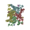
 Movie
Movie Controller


+ Open data

- Basic information
| Entry | Database: PDB / ID: 6jh6 | ||||||
|---|---|---|---|---|---|---|---|
| Title | Structure of RyR2 (F/A/Ca2+ dataset) | ||||||
 Components Components |
| ||||||
 Keywords Keywords | MEMBRANE PROTEIN / cryo-EM | ||||||
| Function / homology |  Function and homology information Function and homology information: / negative regulation of calcium-mediated signaling / negative regulation of insulin secretion involved in cellular response to glucose stimulus / neuronal action potential propagation / insulin secretion involved in cellular response to glucose stimulus / negative regulation of release of sequestered calcium ion into cytosol / response to redox state / negative regulation of heart rate / 'de novo' protein folding / FK506 binding ...: / negative regulation of calcium-mediated signaling / negative regulation of insulin secretion involved in cellular response to glucose stimulus / neuronal action potential propagation / insulin secretion involved in cellular response to glucose stimulus / negative regulation of release of sequestered calcium ion into cytosol / response to redox state / negative regulation of heart rate / 'de novo' protein folding / FK506 binding / smooth muscle contraction / T cell proliferation / calcium channel inhibitor activity / regulation of release of sequestered calcium ion into cytosol by sarcoplasmic reticulum / Ion homeostasis / release of sequestered calcium ion into cytosol / regulation of cardiac muscle contraction by regulation of the release of sequestered calcium ion / sarcoplasmic reticulum membrane / calcium channel complex / peptidylprolyl isomerase / peptidyl-prolyl cis-trans isomerase activity / calcium channel regulator activity / calcium-mediated signaling / protein maturation / Stimuli-sensing channels / Z disc / protein refolding / positive regulation of cytosolic calcium ion concentration / transmembrane transporter binding / signaling receptor binding / membrane / cytoplasm Similarity search - Function | ||||||
| Biological species |  Homo sapiens (human) Homo sapiens (human) | ||||||
| Method | ELECTRON MICROSCOPY / single particle reconstruction / cryo EM / Resolution: 4.8 Å | ||||||
 Authors Authors | Chi, X.M. / Gong, D.S. / Ren, K. / Zhou, G.W. / Huang, G.X.Y. / Lei, J.L. / Zhou, Q. / Yan, N. | ||||||
 Citation Citation | Journal: Proc Natl Acad Sci U S A / Year: 2019 Title: Molecular basis for allosteric regulation of the type 2 ryanodine receptor channel gating by key modulators. Authors: Ximin Chi / Deshun Gong / Kang Ren / Gewei Zhou / Gaoxingyu Huang / Jianlin Lei / Qiang Zhou / Nieng Yan /   Abstract: The type 2 ryanodine receptor (RyR2) is responsible for releasing Ca from the sarcoplasmic reticulum of cardiomyocytes, subsequently leading to muscle contraction. Here, we report 4 cryo-electron ...The type 2 ryanodine receptor (RyR2) is responsible for releasing Ca from the sarcoplasmic reticulum of cardiomyocytes, subsequently leading to muscle contraction. Here, we report 4 cryo-electron microscopy (cryo-EM) structures of porcine RyR2 bound to distinct modulators that, together with our published structures, provide mechanistic insight into RyR2 regulation. Ca alone induces a contraction of the central domain that facilitates the dilation of the S6 bundle but is insufficient to open the pore. The small-molecule agonist PCB95 helps Ca to overcome the barrier for opening. FKBP12.6 induces a relaxation of the central domain that decouples it from the S6 bundle, stabilizing RyR2 in a closed state even in the presence of Ca and PCB95. Although the channel is open when PCB95 is replaced by caffeine and adenosine 5'-triphosphate (ATP), neither of the modulators alone can sufficiently counter the antagonistic effect to open the channel. Our study marks an important step toward mechanistic understanding of the sophisticated regulation of this key channel whose aberrant activity engenders life-threatening cardiac disorders. | ||||||
| History |
|
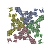
- Structure visualization
| Movie |
Movie viewer |
|---|---|
| Structure viewer | Molecule: MolmilJmol/JSmol |
- Downloads & links
-Download
| PDBx/mmCIF format | 6jh6.cif.gz | 2.4 MB | Display | PDBx/mmCIF format |
|---|---|---|---|---|
| PDB format | pdb6jh6.ent.gz | Display | PDB format | |
| PDBx/mmJSON format | 6jh6.json.gz | Tree view | PDBx/mmJSON format | |
| Others |  Other downloads Other downloads |
-Validation report
| Arichive directory | https://data.pdbj.org/pub/pdb/validation_reports/jh/6jh6ftp://data.pdbj.org/pub/pdb/validation_reports/jh/6jh6 | HTTPS FTP |
|---|
-Related structure data
| Related structure data | 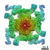 9825MC 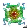 9823C 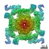 9824C  9826C  6jg3C  6jgzC  6jhnC M: map data used to model this data C: citing same article ( |
|---|---|
| Similar structure data |

-Links
 PDBj
PDBj


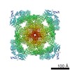
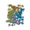
- Assembly
| Deposited unit | 
|
|---|---|
| 1 |
|
-Components
| #1: Protein | Mass: 11798.501 Da / Num. of mol.: 4 Source method: isolated from a genetically manipulated source Source: (gene. exp.) Homo sapiens (human) / Gene: FKBP1B, FKBP12.6, FKBP1L, FKBP9, OTK4 / Production host:  #2: Protein | Mass: 564905.625 Da / Num. of mol.: 4 / Source method: isolated from a natural source / Source: (natural) #3: Chemical | ChemComp-ATP /   Mass: 507.181 Da / Num. of mol.: 4 / Source method: obtained synthetically / Formula: C10H16N5O13P3 / Comment: ATP, energy-carrying molecule*YM Mass: 507.181 Da / Num. of mol.: 4 / Source method: obtained synthetically / Formula: C10H16N5O13P3 / Comment: ATP, energy-carrying molecule*YM#4: Chemical | ChemComp-ZN /   Mass: 65.409 Da / Num. of mol.: 4 / Source method: obtained synthetically / Formula: Zn Mass: 65.409 Da / Num. of mol.: 4 / Source method: obtained synthetically / Formula: Zn |
|---|
-Experimental details
-Experiment
| Experiment | Method: ELECTRON MICROSCOPY |
|---|---|
| EM experiment | Aggregation state: PARTICLE / 3D reconstruction method: single particle reconstruction |
- Sample preparation
| Component | Name: RyR2 in complex with FKBP12.6 / Type: COMPLEX / Entity ID: #1-#2 / Source: NATURAL |
|---|---|
| Source (natural) | Organism: |
| Buffer solution | pH: 7.4 |
| Specimen | Embedding applied: NO / Shadowing applied: NO / Staining applied: NO / Vitrification applied: YES |
| Vitrification | Cryogen name: ETHANE |
- Electron microscopy imaging
| Experimental equipment |  Model: Titan Krios / Image courtesy: FEI Company |
|---|---|
| Microscopy | Model: FEI TITAN KRIOS |
| Electron gun | Electron source:  FIELD EMISSION GUN / Accelerating voltage: 300 kV / Illumination mode: FLOOD BEAM FIELD EMISSION GUN / Accelerating voltage: 300 kV / Illumination mode: FLOOD BEAM |
| Electron lens | Mode: BRIGHT FIELD |
| Image recording | Electron dose: 48.6 e/Å2 / Film or detector model: GATAN K2 SUMMIT (4k x 4k) |
- Processing
| EM software |
| ||||||||||||||||
|---|---|---|---|---|---|---|---|---|---|---|---|---|---|---|---|---|---|
| CTF correction | Type: PHASE FLIPPING AND AMPLITUDE CORRECTION | ||||||||||||||||
| Symmetry | Point symmetry: C4 (4 fold cyclic) | ||||||||||||||||
| 3D reconstruction | Resolution: 4.8 Å / Resolution method: FSC 0.143 CUT-OFF / Num. of particles: 44288 / Symmetry type: POINT |
